- Company
- Products
- Catalogs
- News & Trends
- Exhibitions
OSE-2800
1 /2Pages
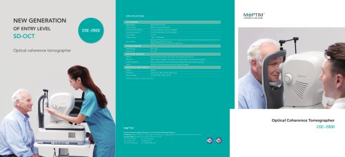
OSE-2800
1 /2Pages
Catalog excerpts

Optical source Axial resolution (optical) 5 microns (optical), 3.6 microns (digital) Transverse resolution 15 microns (optical), 3 microns (digital) A-scan depth Diopter range Scan patterns Macular: HD line scan (6 mm or 12 mm), Cube scan (6 mm x 6 mm), Six-line radial scan, Multi (X-Y: 5 x 5) Disc: Cube scan (6 mm x 6 mm) Anterior: HD line scan (6 mm), 6-line radial scan FUNDUS IMAGING Optical coherence tomographer SOFTWARE ANALYSIS Macula Retina thickness analysis; 3D view; En-face analysis; EDI function RNFL analysis; Ganglion cell analysis; Cup-disk analysis; OU comparative analysis Anterior Segment Manual measurement; Corneal thickness analysis; Epithelial thinckness analysis DICOM conformance; Remote viewer software available ELECTRICAL AND PHYSICAL Weight Source voltage Specifications subject to change without notice. Optical Coherence Tomographer OSE-2800 Shenzhen Moptim Imaging Technique Co., Ltd. (A Certainn Technology Company) Headquarter: Bldg. 2-C, Section 2, GOTO Digital Technology Park, Longgang District, No.137 Bulan Rd., Shenzhen 518112, China European Office: Evelpidon 61-63, 4th Floor, Athens P.O 11362 Tel: +30 210 5750572
Open the catalog to page 1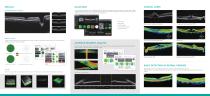
High definition OCT imaging For comprehensive glaucoma analysis, OSE-2800 SD-OCT offers two scan patterns, glaucoma cube scan in macular area for RNFL analysis and glaucoma cube scan in disc area for ONH analysis. Evenly distributed sampling point with 500 x 200 A-scans provides reliable information for early glaucoma detection and management. CLINICAL CASES · GCC analysis · RNFL analysis Retinal pigment epithelium detachment Macular hole Vitreomacular traction syndrome (VMT) Central serous chorioretinopathy (CSC) · Cup-disc analysis Polypoidal choroidal vasculophy (PCV) · OU comparative analysis...
Open the catalog to page 2All Moptim catalogs and technical brochures
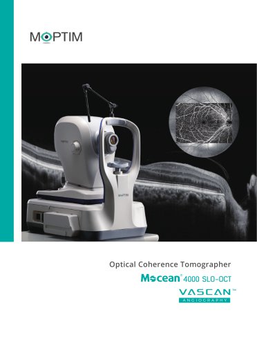 Mocean™ 4000
8 Pages
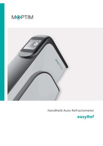 easyRef
4 Pages
 SL-M6
SL-M65 Pages
 OPM 500
2 Pages
 DEA
DEA8 Pages
Archived catalogs
 iRef
iRef3 Pages
 Mocean 4000
8 Pages
 Colombo IOL Optical Biometer
3 Pages
 MOPTIM catalogue 2022
12 Pages
- Fixed ophthalmic examination
- Software module
- Laboratory refractometer
- Hand-held ophthalmic examination instrument
- Slit lamp
- Digital refractometer
- Table slit lamp
- Ophthalmoscope
- Operating microscope
- Portable refractometer
- Analysis software module
- Refractometer ophthalmic examination
- Automatic refractometer
- Medical imaging software module
- Ophthalmic biometer
- Visualization software module
- OCT ophthalmoscope
- Tablet PC software module
- Retinal imaging instrument
- Dry eye diagnosis system