
Catalog excerpts
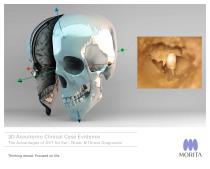
3D Accuitomo Clinical Case Evidence The Advantages of DVT for Ear-, Nose- & Throat-Diagnostic Thinking ahead. Focused on life.
Open the catalog to page 1
Dear Colleagues, I am very happy to present you now some data on cone beam tomography (digital volume tomography) with this booklet. This imaging procedure is highly interesting in otorhinolaryngology and I am convinced that it will play a major role in future routine diagnosis. In order to summarize detailed knowledge on this procedure we decided to create this booklet. Special thanks to my co-worker Dr. Christian Güldner and of course also to Morita Company that nally made this booklet possible. Please inform yourself of the modern technique. It does not only on the achievements of the...
Open the catalog to page 2
[04 - 05] Introduction of 3D Accuitomo - Compact, High-Resolution, Low-Dose [06 - 09] Efficient Workflow Integration of DVT [10 - 11] i-Dixel Image Processing Software Temporal Bone Cases [12 -13] Anatomy of the Temporal Bone in Digital Volume Tomography [14-15] Axial Plain, caudal [16-17] Axial Plain, cranial [18 -19] Coronal Plain, from anterior to posterior [20 - 21] Sagittal Plain, from lateral to medial [22 - 23] Osteoma of temporal bone Anterior Skull Base Cases [24 - 25] Anatomy of the Anterior Skull Base in Digital VolumeTomography [26 - 30] Coronal Plain, from anterior to posterior...
Open the catalog to page 3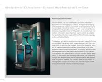
Introduction of 3D Accuitomo – Compact, High-Resolution, Low-Dose Advantages of Cone Beam 3D Accuitomo 170 is a cone-beam CT or also called DVT; digital volume tomography, which is designed for imaging of head-and-neck regions, especially for temporal bone and sinuses. With one single rotation of the x-ray tube and area detector around the head of the patient, it provides high-resolution volumetric data of a small region, which is suitable for specialized medical applications of otorhinolaryngology and maxillofacial elds. The patient is in sitting position during scan, instead of lying on...
Open the catalog to page 4
This system is also designed to be space-efcient compared to existing CT systems in the market, it is even suitable for a small clinic. This type of device can be a far more practical and useful tool than an existing CT for special applications requiring detailed imaging of the ne bone structures of the middle ear, etc. By using an area detector with very small pixels, it is possible to obtain high-resolution images, compared to a medical CT. Because of the two dimensional detector, the resolution of the sagittal and coronal slices is as high as the one of the axial slices. The volume data...
Open the catalog to page 5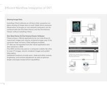
Efcient Workow Integration of DVT Sharing Image Data Installing i-Dixel software on all intra-clinic computers enables sharing of image data on each linked client computer. Observation of images on non-network computers can be achieved with the One Data Viewer, and the One Volume Viewer without installing i-Dixel. 3D Accuitomo 170 One Data Viewer & One Volume Viewer Software These unique J. Morita applications let you view three dimensional images and volume rendered images even if the computer does not have i-Dixel software installed. CT data can be exported from the i-Dixel application...
Open the catalog to page 6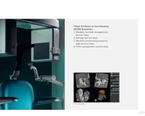
i-Dixel Conforms to the Following DICOM Standards: 1. Modality worklist management service class 2. Storage service class 3. Modality performed procedure step service class 4. Print management service class
Open the catalog to page 7
Efcient Workow Integration of DVT i-Dixel Image Processing Software It can be used as a database to archive a wide variety of image information. Its multiple image processing functions can easily access and manipulate many types of information for 2D and 3D images. Volume Rendering Volume rendering of CT data produces three dimensional images. Select the area of interest and adjust the controls for the histogram to create a detailed image of very ne structures. Real Time Re-Slice Slices and volume rendered images can be linked and easily manipulated in real time. Curved MPR (cMPR) This way...
Open the catalog to page 8
Report Comments • XYZ view windows • Re-slice • Zoom • Rotate • Histogram • Edge Enhancement • Distance and Angle Measurement • Negative Image • Mirror Image • Slice Distance Measurement • Surface Rendering • DICOM 3.0 Compatible • Brightness Conversion • Spatial Frequency Filter • Patient Orientation Display • Density Measurement
Open the catalog to page 9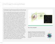
i-Dixel Image Processing Software The Multiple Image Processing Functions and Easy Access The arm rotates 360° around the center of the exposure region in 18 seconds (Standard Mode) as the x-ray head emits a cone-shaped beam. The multiple projections created during the arm’s rotation are converted to a digital signal by the at panel detector and transmitted to the computer. After any necessary supplemental or corrective processing, the digital information is converted into a three dimensional CT image using an image reconstruction algorithm, and a high resolution image appears on the...
Open the catalog to page 10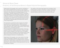
Temporal Bone Cases Anatomy of the Temporal Bone in Digital Volume Tomography Digital Volume Tomography can be used for the display of the complex threedimensional anatomy in the temporal bone very well. This is due to the fact that DVT enables high resolution, low artifacts and 3 orthogonal plains in one examination. Possible indications for the DVT in temporal bone diagnostics can be inammatory diseases, control of middle ear prosthesis and cochlear implants as well as the evaluation of the inner ear itself. The absolute precondition of successful use of DVT in the lateral skull base are...
Open the catalog to page 12
The favourable projections are summarised in the following table:
Open the catalog to page 13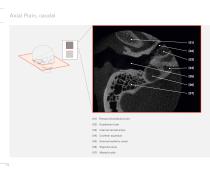
Axial Plain, caudal Internal carotid artery Cochlear aqueduct External auditory canal Sigmoid sinus Eustachian tube Mastoid cells
Open the catalog to page 14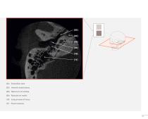
Eustachian tube Internal carotid artery Basal turn of cochlea Manubrium mallei Long process of incus Facial recessus
Open the catalog to page 15
Axial Plain, cranial [08] Basal turn of cochlea [10] Long process of incus [12] Apical turn of cochlea [13] Middle turn of cochlea Internal auditory canal Malleus neck
Open the catalog to page 16All Morita catalogs and technical brochures
-
 Tri Auto ZX2+
8 Pages
-
 Instrument brochure
16 Pages
-
 EndoWave Feilen
2 Pages
-
 Signo T100
12 Pages
-
 Signo Z300
12 Pages
-
 Signo T500
13 Pages
-
 Signo G10 II
41 Pages
-
 Root ZX mini
2 Pages
-
 3D Accuitomo 170
16 Pages
-
 Veraviewepocs 2D
10 Pages
-
 Veraview IC5 HD
5 Pages
-
 GUMMETAL®
5 Pages
-
 Lubrina 2
8 Pages
-
 Veraviewepocs 3D R100
16 Pages
-
 AdvErL EVO
6 Pages
-
 TriAuto mini
10 Pages
-
 Implant Dentistry
36 Pages
-
 Veraview X800
10 Pages
-
 TorqTech handpieces
2 Pages
-
 Veraview iX
2 Pages
-
 EndoWave OTR Sequence FM
2 Pages
-
 Genuine Root ZX
2 Pages
-
 i-Dixel Web
2 Pages

Archived catalogs
-
 Soaric for endodontics
2 Pages
-
 Soaric
19 Pages
-
 PenCure 2000
2 Pages
-
 Brochure "Endodontics is an Art"
10 Pages
