- Company
- Products
- Catalogs
- News & Trends
- Exhibitions
Implant Dentistry
1 /36Pages

Implant Dentistry
1 /36Pages
Catalog excerpts

Implant Dentistry The Use of Cone Beam Computed Tomography (CBCT) Thinking ahead. Focused on life.
Open the catalog to page 1
Prof. Dr. med. dent. Michael Bornstein Prof. Dr. med. dent. Daniel Buser Dental degree and thesis at the University of Basel. Board-certified oral surgeon since June 2003. In 2004, visiting assistant professor at the Department of Periodontics (Prof. Dr. D. Cochran) at the University of Texas Health Science Center at San Antonio, USA. Since autumn 2007 head Section of Dental Radiology and Stomatology at the Department of Oral Surgery and Stomatology at the University of Bern. In October 2009, Habilitation (Privatdozent / PD) in the field of „Oral Surgery and Stomatology“. Since 2014, Associate...
Open the catalog to page 2
Technical details of the 3D Accuitomo 170 6 Dose optimization for CBCT imaging 8 CBCT imaging for implant treatment planning 10 in the anterior maxilla Case example: Replacement of a maxillary 12 central incisor following dento-alveolar trauma CBCT imaging for implant treatment planning 14 in the posterior maxilla Case example: Replacement of a missing first 16 left maxillary molar following tooth loss due to endodontic complications Case example: Replacement of missing maxillary 18 premolars and molars in the posterior right maxilla (distal extension situation) CBCT imaging...
Open the catalog to page 3
1. Harris D, Horner K, Gröndahl K, Jacobs R, Helmrot E, Benic GI, Bornstein MM, Dawood A, Quirynen M: Guidelines for the use of diagnostic imaging in implant dentistry 2011: update of the E.A.O. A consensus workshop organized by the European Association for Osseointegration in the Medical University of Warsaw, Poland. Clin Oral Implants Res 2012;23:1243-53. 2. Mozzo P, Procacci C, Tacconi A, Tinazzi Martini P, Bergamo Andreis IA. A new volumetric CT machine for dental imaging based on the cone-beam technique: preliminary results. Eur Radiol 1998;8:1558-64. 4. Guerrero ME, Jacobs R, Loubele M,...
Open the catalog to page 4
Preface Proper diagnosis and treatment planning in implant dentistry is done with a clinical examination and appropriate radiographs. For many years, the information required to satisfy these goals has been obtained from clinical examination and most commonly two-dimensional (2D) imaging such as intraoral periapical, lateral cephalometric and panoramic radiography. Using these imaging modalities, implants have been used predictably and with high success rates in clinical practice for more than 30 years. The decision to proceed to three-dimensional (3D) imaging should be based on clearly identified...
Open the catalog to page 5
Technical Details of the 3D Accuitomo 170 The 3D Accuitomo 170 is the 4th generation of the Accuitomo product line. It combines a high level of image quality with efforts to minimize patient dosage. The device offers different imaging modes, Fields of View (FOV) varying from small to large (maxillo-facial) FOVs, voxel sizes from 80 μm to 250 μm, zoom reconstructions to analyze areas of special interest with voxel sizes as small as 80 μm, and a DICOM 3.0 compliant imaging software (i-Dixel 3D). Image acquisition with the 3D Accuitomo 170 is done by a rotating gantry composed of an X-ray source...
Open the catalog to page 6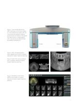
Figure 1: The principle behind the CBCT technique, as its name implies, is a cone-shaped X-ray beam with the X-ray source and detector rotating around a point of interest in the patient. This figure shows a rotating gantry with x-ray source (so-called emitter) and detector attached to it. Emitter Figure 2 (left): Orthogonal planes (axial, sagittal and coronal) in a patient referred for implant treatment planning. Figure 3 (right): 3D volume rendered jaw in a patient referred for dental implant treatment planning. Figure 4: Multiplanar reformation (MPR) of the maxilla in a patient referred for...
Open the catalog to page 7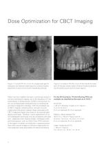
Dose Optimization for CBCT Imaging Figure 1: A small FOV (4 x 4 cm) of a single-tooth gap for evaluation and treatment planning prior to dental implant placement in area of the first left mandibular premolar. Figure 2: A medium FOV (8 x 4 cm) of the maxilla for evaluation of the peri-implant status of dental implants placed at the left lateral incisor and first molar regions. Patient risk from radiation has been a continuing concern in oral and maxillofacial imaging, due to the frequency of X-ray examinations in dental practice. ALARA is the acronym for As Low As Reasonably Achievable and is...
Open the catalog to page 8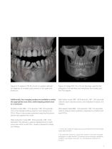
Figure 3: A medium FOV (8 x 8 cm) in a patient referred for diagnosis of multiple cystic lesions in the upper and lower jaw. Figure 4: A large FOV (14 x 10 cm) has been used for the evaluation of all maxillary and mandibular third molars and the TMJ regions. Additionally, four imaging modes are available to select the appropriate scan time, while keeping patient dose to a minimum: High fidelity mode (360˚: 30.8 seconds / 180˚: 15.8 seconds): Used for zoom reconstructions, and indicated for adults, not children. Standard mode (360˚: 17.5 seconds / 180˚: 9.0 seconds): This is the standard setting...
Open the catalog to page 9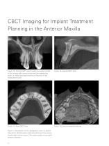
CBCT Imaging for Implant Treatment Planning in the Anterior Maxilla Figure 1A: Coronal CBCT slice through the alveolar socket of the missing right central incisor and the neighboring teeth. (*) Nasal openings (foramina of Stenson) of the nasopalatine canal Figure 1B: Sagittal CBCT slice Figure 1C: Axial CBCT slice Figure 1D: Volume rendered data set Figure 1: Visualization of the nasopalatine canal in a patient referred for dental implant treatment planning in the anterior maxilla (right central incisor). The canal consists of two parallel bony channels. 10
Open the catalog to page 10
Relevant clinical questions Is a correct 3-dimensional (3D) implant positioning possible? Besides reestablishing function, the esthetic result of the implant rehabilitation is of great importance in the anterior maxilla. Therefore, the clinician needs to achieve a correct oro-facial, mesio-distal, and corono-apical position of the prospective implant shoulder. Can we achieve good primary stability of the implant in the desired position? If primary stability of an inserted dental implant is not achievable, the clinician has to decide between site development using guided bone regeneration (GBR)...
Open the catalog to page 11All Morita catalogs and technical brochures
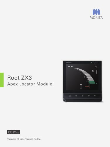 Root ZX3 - Apex Locator
2 Pages
 Tri Auto ZX2+
8 Pages
 Instrument brochure
16 Pages
 EndoWave Feilen
2 Pages
 Signo T100
12 Pages
 Signo Z300
12 Pages
 Veraview iX
2 Pages
 Signo T500
13 Pages
 Signo G10 II
41 Pages
 Root ZX mini
2 Pages
 3D Accuitomo 170
16 Pages
 Veraviewepocs 2D
10 Pages
 TriAuto mini
10 Pages
 Veraview IC5 HD
5 Pages
 Veraviewepocs 3D R100
16 Pages
 GUMMETAL®
5 Pages
 Lubrina 2
8 Pages
 AdvErL EVO
6 Pages
 Veraview X800
10 Pages
 TorqTech handpieces
2 Pages
 EndoWave OTR Sequence FM
2 Pages
 Genuine Root ZX
2 Pages
 i-Dixel Web
2 Pages

- Anatomy model
- Training anatomy model
- Trolley-mounted laser
- Dental treatment unit
- Visualization software
- Radiology software
- Dental unit with light
- Dental model
- Drill
- Micromotor
- Diagnostic software
- Electric micromotor
- Turbine
- Dental radiography system
- Dental turbine
- Air turbine
- Dental unit with LED light
- Dental micromotor
- Data management software
- Dental unit with hygiene system