- Company
- Products
- Catalogs
- News & Trends
- Exhibitions
Veraviewepocs 3D R100
1 /16Pages

Veraviewepocs 3D R100
1 /16Pages
Catalog excerpts

Thinking ahead. Focused on life.
Open the catalog to page 1
Veraviewepocs 3D R100 MORITA image excellence for all offices The Veraviewepocs 3D R100 offers the unique MORITA image quality for every dental Practice. The Veraviewepocs 3D R100 has revolutionized 3D imaging and continues to set standards. Superior image quality in 3D and 2D, the MORITA-exclusive Panoramic Scout function and the MORITA-exclusive Reuleaux image format are just a few examples. In addition, there are features such as 6 selectable exposure areas, an automatic exposure for panorama shots and innovative techniques for automatic dose reduction.
Open the catalog to page 2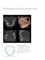
3D Reuleaux Full Arch Field of View Blue line indicates full arch FOV, equivalent to ∅ 10 0 mm. Patent Pending Technology Morita's completely unique 3D Reuleaux Full Arch FOV abandons the typical cylinder with a convex triangle shape. By more closely matching the natural dental arch form, this groundbreaking FOV reduces dose by excluding areas outside the region of interest and allows a complete scan of the maxilla and/or the mandible. Reduce dose with the innovative 3D Reuleaux FOV. 4
Open the catalog to page 4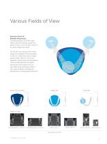
Various Fields of View Exposure Areas for Multiple Diagnostics The Veraviewepocs 3D R100 model offers a total of 6 exposure areas from Ø 40 x H 40 mm up to Ø 100 x H 80 mm for various diagnostic needs. The full arch scan captures the maxilla and/or the mandible with the equivalent of 100 mm in diameter and two height options of 50 or 80 mm. Its full arch capability, reduced dose, and exceptional clarity are ideal features for implant planning and oral surgery. This unit also offers small and medium field of view sizes suitable for endodontics, periodontics, as well as general dentistry.
Open the catalog to page 5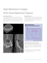
High Resolution Images With Dose Reduction Feature Dose Reduction Feature Through advanced engineering, a Dose Reduction Mode optimizes the intensity of the X-rays which lowers exposure for easily penetrated tissues. Dose is reduced to a mere 60% of the standard mode.* By maximizing efficiency, the maxillary sinus membrane appears sharper than ever before with fewer artifacts.** Resolution & Clarity Veraviewepocs offers high resolution images of 125 µm voxel. It provides clear images of the periodontal pocket, the periodontal ligament, and the alveolar bone. It is extremely useful for implant...
Open the catalog to page 6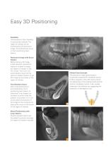
Flexibility Veraviewepocs offers flexibility in positioning methods. The region of interest can be positioned by the panoramic image, the bi-directional scout, or the 5 positioning laser beams. Panoramic Image with Scout Feature Before taking a 3D image, a high resolution panoramic exposure is taken to target the region of interest on the PC monitor. The C-arm will automatically move into the optimum patient position to get 3D images at the center of the region of interest. Two-Direction Scout After initial positioning is accomplished by the 3 positioning laser beams, bidirectional X-ray images...
Open the catalog to page 7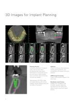
Planning Process Successful placement of implants starts with the very critical and detailed planning process. Identification of structures such as the sinus cavity, inferior alveolar nerve, and clear views of the bone structure are needed. Veraviewepocs 3D R100 is ideal for implant planning with full arch imaging, industry leading clarity, and low dose to the patient. Software i-Dixel 2.0 software offers advanced implant planning features, plus compatibility with popular third party software. cMPR Image Processing Create cross sectional images of the dental arch. Mandibular Canal Tracing Highlight...
Open the catalog to page 8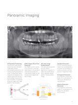
AF Automatic Positioning This function makes patient positioning nearly effortless. A light beam sensor automatically positions the unit without requiring the patient to move. The light beam sensor measures the distance to the patient's teeth, then the arm automatically moves into the optimal position. This process produces images with a high degree of reproducibility. DDAE (Digital Direct Auto Exposure) The DDAE function controls X-ray emission in real time depending on the area being examined and produces a wide dynamic range, as well as sharp and exceptionally clear images. AIE (Auto Image...
Open the catalog to page 9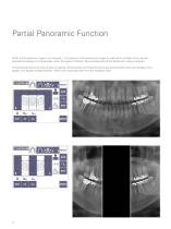
Partial Panoramic Function When a full panoramic image is not required, 1 to 5 sections of the panoramic image, as well as the maxillary sinus, can be excluded to expose only those areas within the region of interest. By excluding parts of the dental arch, dose is reduced. The partial panoramic function is easy to operate. Simply press the Partial Panorama key and the panoramic and maxillary sinus appear with equally divided sections. Select any to exclude them from the irradiation area.
Open the catalog to page 10
High Speed The Veraviewepocs system offers high speed performance requiring only 2.6 to 5.8 seconds for a lateral projection. The speed helps ensure high quality images each and every time. For pediatric patients, the reduced scan time is especially helpful as repeat images due to patient movement are virtually eliminated. Low Dose With only a tenth of the dose compared to a conventional X-ray*, the exposure level is significantly reduced. High Quality Image with Wide Dynamic Range You obtain far more information about hard and soft tissue - with just a single acquisition. Variable Imaging Processing The...
Open the catalog to page 11
Clinical Cases Implantology The patient was seen for a routine follow-up visit following implant placement in the area of the left maxillary lateral incisor. The implant had been placed 3 months earlier. The coronal, sagittal, and axial planes revealed a large, round, well defined, non-corticated, low density area associated with the apical aspect of the implant. The high resolution images also shows absence of the buccal cortical plate confirming a poor prognosis for the case due to peri-implantitis. Endodontics The patient reported history of trauma in the left anterior maxilla. A cone beam...
Open the catalog to page 12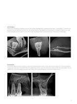
Oral Surgery Patient was referred for surgical removal of the mesial impacted right mandibular third molar. A cone beam CT volume was acquired with the 3D R100 to determine root-nerve proximity. The sagittal and coronal views revealed the path of the right inferior alveolar canal through the roots of the right mandibular third molar and thinning of the lingual cortical plate. Periodontics The patient reported tooth sensitivity in the left maxillary second molar. A small volume cone beam CT of the left posterior maxilla was acquired with the 3D R100. The sagittal and coronal views showed severe...
Open the catalog to page 13All Morita catalogs and technical brochures
 Root ZX3 - Apex Locator
2 Pages
 Tri Auto ZX2+
8 Pages
 Instrument brochure
16 Pages
 EndoWave Feilen
2 Pages
 Signo T100
12 Pages
 Signo Z300
12 Pages
 Veraview iX
2 Pages
 Signo T500
13 Pages
 Signo G10 II
41 Pages
 Root ZX mini
2 Pages
 3D Accuitomo 170
16 Pages
 Veraviewepocs 2D
10 Pages
 TriAuto mini
10 Pages
 Veraview IC5 HD
5 Pages
 GUMMETAL®
5 Pages
 Lubrina 2
8 Pages
 AdvErL EVO
6 Pages
 Veraview X800
10 Pages
 Implant Dentistry
36 Pages
 TorqTech handpieces
2 Pages
 EndoWave OTR Sequence FM
2 Pages
 Genuine Root ZX
2 Pages
 i-Dixel Web
2 Pages

- Anatomy model
- Training anatomy model
- Trolley-mounted laser
- Dental treatment unit
- Visualization software
- Radiology software
- Dental unit with light
- Dental model
- Drill
- Micromotor
- Diagnostic software
- Electric micromotor
- Turbine
- Dental radiography system
- Dental turbine
- Air turbine
- Dental unit with LED light
- Dental micromotor
- Data management software
- Dental unit with hygiene system