- Company
- Products
- Catalogs
- News & Trends
- Exhibitions
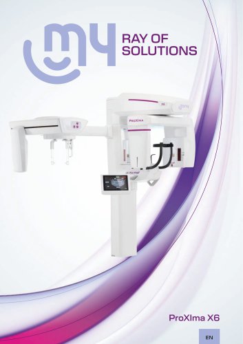
ProXIma X6 | MyRay
1 /17Pages
ProXIma X6 | MyRay
1 /17Pages
Catalog excerpts
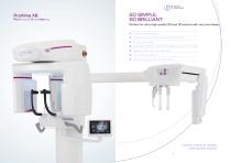
Professional X-ray Imaging SO SIMPLE, SO BRILLIANT Perfect for ultra-high quality 2D and 3D exams with very low doses. Modern minimal design System can easily be integrated with CEPH arm Extremely compact Ultra-high resolution 2D and 3D images that are rich in detail Effective, safe, real-time diagnoses User-friendly software Enhanced communication with patient Intuitive, practical, reliable. Nothing else needed. 3
Open the catalog to page 2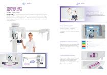
TAKES SHAPE AROUND YOU AIRgonomics version Flexible configuration An exclusive wall-mounted installation without any floor obstacles not only saves space but also facilitates access for patients. ProXIma X6, lets you choose from among several different configurations to capture 2D, 3D and CEPH images. If desired, new functions can be added at a later stage. To adapt perfectly to the available space, the control panel is positioned according to your usage preferences, while the ceph arm can be installed both on the left or right of the column. Relax lighting system Gives your practice a distinctive...
Open the catalog to page 3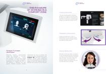
THE PLEASURE OF WORKING IN A COMFORT ZONE Virtual control panel The user-friendly graphic interface guides you through the process step by step: from selection of the exam to execution of the scan, providing direct access to all device functions via PC. Greatly increases the number of examinations you can perform each working day, ensuring images remain accurate and high-quality. Full-touch 7” on-board control panel Featuring modern, ultra-compact design, the integrated 7” full-touch control panel guides you - simply and intuitively - through every stage of positioning and image acquisition....
Open the catalog to page 4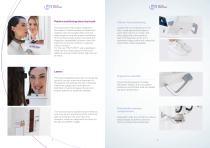
Patient positioning/securing tools Patient foot positioning The ergonomic head support adapts to the shape of individual patients’ heads and, together with the supplied bites and subnasal supports, ensures proper positioning of the arches, a high-quality final result and diagnostic repeatability of scans, even with edentulous persons, children or patients without incisors. For the new “15x11 DENT” scan, dedicated head and sub-nasal supports have been added to ensure proper patient alignment at all times. A laser beam is projected onto the floor, remaining correctly aligned even if the column...
Open the catalog to page 5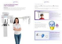
YOUR PATIENTS FIRST AND FOREMOST ProXIma X6 has been designed to reduce X-ray emissions while maintaining ultra-high image quality. This is possible thanks to automatisms, functions and accessories that calibrate X-ray doses according to the patient’s actual needs and their anatomy, protecting the most sensitive areas. Cutting-edge protocols Available for both 2D (QuickPAN and QuickCEPH) and 3D (QuickSCAN) examinations, these provide accurate images but with lower doses than standard acquisitions. These are particularly useful for post-surgical follow-ups or identifying any macrostructures, such...
Open the catalog to page 6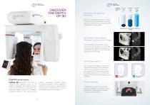
Optimised 3D scanning protocols image quality MINIMUM EXPOSURE TIME Each FOV has three execution modes to adapt to all clinical needs, ensuring exams are performed according to real needs with extreme ease. QUICKSCAN STANDARD SUPER HD MODE 3D SMART (Streak Metal Artifacts Reduction Technology) Automatically ensures anatomical structures remain sharp even where there are metal objects (amalgam or implants) that might compromise the quality of the 3D image. Scout View system By viewing two images of the patient, one lateral and one frontal obtained with a very low radiation dose, you can align...
Open the catalog to page 7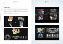
3D dental exams Sectoral tomographic images of complete or partial dentition, individual arches, maxillary or mandibular or both, including the upper airways (nose, throat, sinuses). The introduction of the extended “15x11 DENT” FOV also makes analysis more versatile and is able to capture both dental arches and part of the temporomandibular joint on adult patients. • Typical FOVs for dental scans on adult or paediatric patients: 6x6, 8x6, 8x8, 11x6, 11x8, 11x11, 15x11 DENT
Open the catalog to page 8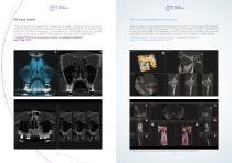
3D temporomandibular joint exams Three-dimensional images of the maxillary sinus region, including nose and a portion of the cheekbone area or the maxillary sinuses area depending on the patient’s build. Useful for verifying morphology or anomalies and pathologies such as sinusitis, tumours, obstructions, genetic malformations, opening of the middle meatus. Ability to capture both temporomandibular joints, verify the morphology of the relative bone structures, diagnose fractures or traumas and assess condylar translation to study joint functionality. The available set of FOVs allows for acquisition...
Open the catalog to page 9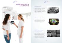
2D IMAGING THAT’S A MUST-HAVE MultiPAN function With just a single scan - and a dose equal to that of a single traditional panoramic X-ray - 5 different focus layers can be obtained. You can then select the one that best highlights the diagnostic detail of interest. iPAN function (Focus-Free) Lets you obtain a single panoramic image automatically by merging the layers generated by the MultiPAN function and selecting the most infocus portions of each of them. 2D PiE (Picture image Enhancer) filters on PAN Focus-Free function These automatically optimise each layer captured with the MultiPAN function...
Open the catalog to page 10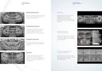
Standard Panoramic Allows a complete, accurate view of the dental arches, maxillary sinuses and temporomandibular joints. Optimised collimated interproximal projection with a low dose to investigate dental crowns. An alternative to intraoral bitewings, with a less invasive and more comfortable procedure. Orthogonal panoramic Compared to a standard panoramic image, this highlights interproximal spaces perfectly; the entire root structure is free from any overlapping. Maxillary Sinuses (frontal and lateral) Paediatric panoramic Creates an image that allows dentists to assess the health of the maxillary...
Open the catalog to page 11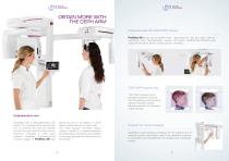
OBTAIN MORE WITH THE CEPH ARM Repositionable 2D PAN/CEPH sensor ProXIma X6 allows you to perform both repositioned in the two slots used for panoramic and cephalometric exams 2D exams. Outstanding efficiency and using the the same sensor, which can be versatility. TOP CEPH positioning TOP CEPH positioning for paediatric patients reduces thyroid exposure and prevents sensor-shoulder contact, allowing inclusion, if necessary, of the skullcap. Cephalometric arm Equipped with a latest-generation 2D sensor, the cephalometric examination arm is compact and can be installed on both the right and left...
Open the catalog to page 12All MyRay catalogs and technical brochures
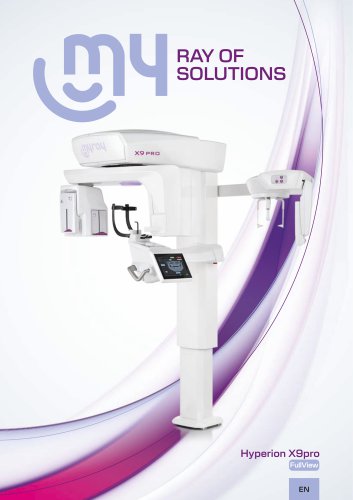 Hyperion X9 Pro FullView
13 Pages
 Software Neowise
7 Pages
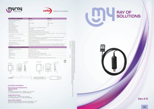 Zen-X E | MyRay
4 Pages
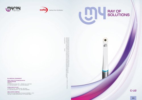 C-U2 | MyRay
4 Pages
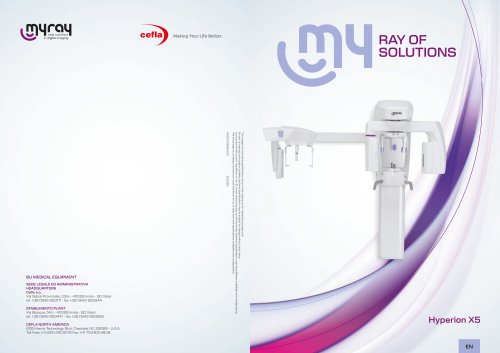 Hyperion X5 | MyRay
18 Pages
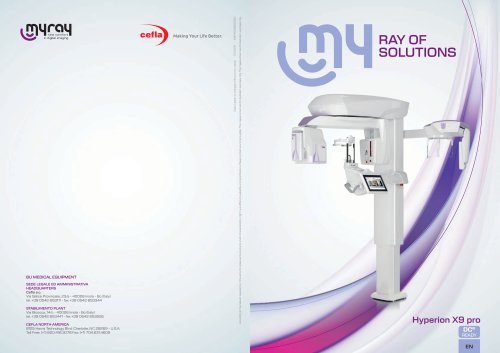 Hyperion X9 Pro | MyRay
18 Pages
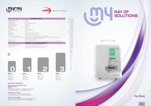 Hy-Scan | MyRay
4 Pages
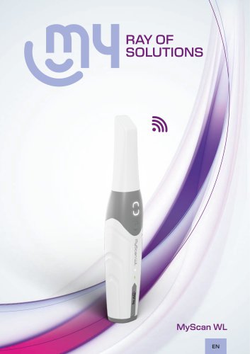 MyScan WL | MyRay
7 Pages
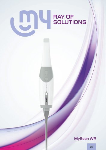 MyScan WR | MyRay
7 Pages
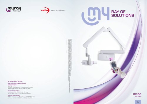 RX DC eXTend | MyRay
4 Pages
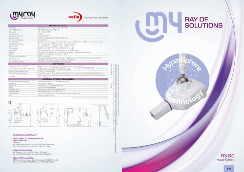 RX DC HyperSphere | MyRay
4 Pages
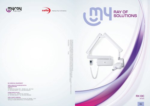 RX DC | MyRay
4 Pages
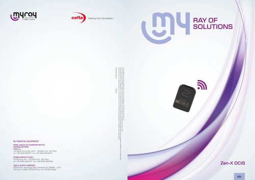 Zen-X DCiS | MyRay
6 Pages
- Analysis software
- Visualization software
- Radiology software
- Tablet computer software
- Tablet PC software
- Flat panel detector
- Monitoring software
- Diagnostic software
- Planning software
- Cloud-based software
- Portable flat panel detector
- Dental software
- Dental radiography system
- Data management software
- AI-assisted software
- Artificial intelligence software
- Surgery software
- Digital dental radiography system
- 3D scanner
- Simulation software