5G XL (MED)
1 /15Pages

5G XL (MED)
1 /15Pages
Catalog excerpts

CONE BEAM 3D IMAGING
Open the catalog to page 1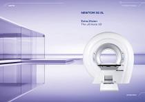
CONE BEAM 3D IMAGING BEYOND VISION
Open the catalog to page 2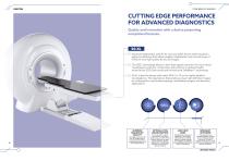
CONE BEAM 3D IMAGING CUTTING EDGE PERFORMANCE FOR ADVANCED DIAGNOSTICS Quality and innovation with a device presenting exceptional features. 5G XL O Advanced diagnostics with 5G XL, the only CBCT device with lying down patient positioning that offers excellent stabilisation and a broad range of FOVs for very high quality 3D and 2D images. □ The CBCT technology allows to have high spatial resolution for bone tissue investigations with low X-Ray dose. Special focus on patient health, enhanced by ECO Dose mode and the exclusive SafeBeam™ technology. □ 5G XL is the first device with native FOV 21...
Open the catalog to page 3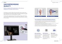
CONE BEAM 3D IMAGING UNCOMPROMISING QUALITY Superior standard 3D examinations with a device designed for excellent performance. High definition volumetric images of bone tissue with a “native” isotopic voxel, non-overlapping sections and fewer artifacts. With CBCT technology, 5G XL offers faster examinations and low doses of radiation with greater safety for the patient, better performance and an increasingly efficient workflow. The high quality images generated by 5G XL are ideal for multiple medical fields, such as dental-maxillofacial diseases, otorhinolaryngology applications, complete analysis...
Open the catalog to page 4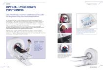
CONE BEAM 3D IMAGING OPTIMAL LYING DOWN POSITIONING User-friendliness, maximum stabilisation and quality for diagnoses using new medical applications. 5G XL is the only CBCT device available on the market with patient in a lying down position. The motor-driven patient table made of carbon fibre, which can be controlled from the on-board console or from the PC, allows to adapt the examination to any image acquisition need with patient lying down in a prone or supine, cranial-caudal or caudal-cranial position. The open gantry facilitates access to the scanning area and eliminates any sensation...
Open the catalog to page 5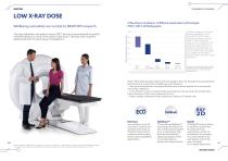
CONE BEAM 3D IMAGING LOW X-RAY DOSE X-Ray Dose comparison of different examination technologies: MSCT, CBCT, 2D Radiography Wellbeing and safety are central to NEWTOM research. The different settings of FOVs and Resolution available in the use of the NEWTOM 5G XL allow a wide range of different screening based on different diagnostic needs. In some configurations, for a preliminary screening exam, for example the 18x16 Standard, it is possible to obtain 3D images with an X-rays dose emitted comparable to two X-Rays 2D radiographs (Antero-Posterior and Latero-Lateral projection)**. Effective Dose...
Open the catalog to page 6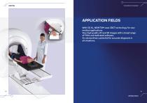
CONE BEAM 3D IMAGING APPLICATION FIELDS With 5G XL, NEWTOM uses CBCT technology for new medical applications. Very high quality 2D and 3D images with a broad range of FOVs and dedicated software. An extraordinary potential for accurate diagnosis in all situations.
Open the catalog to page 7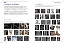
Orthopaedic applications Images generated by 5G XL, with their high resolution and quality, allow an in-depth study of the upper and lower limbs not only to diagnose fractures, dislocations, luxations or misalignment but also to define the bone and joint structure resulting from pathological alterations, to detect small bone fragments and to assess diseases in small joints, even when metal screws are present. Excellent acquisitions that exceed the limitations of CT examinations or those typical of 2D image acquisitions, in which a dedicated visual alignment cannot always prevent overlapping bone...
Open the catalog to page 8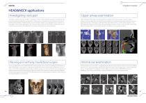
CONE BEAM 3D IMAGING HEAD&NECK applications Investigating neck pain The better spatial resolution of CBCT, compared to MSCT, allows detailed analyses of trabecular and cortical structures to identify any dysplastic, inflammatory, traumatic or micro-traumatic elements. Relationships between vertebral bodies are also perfectly legible, thus highlighting any distortion or subluxation. 3D volumes generated with 5G XL are ideal to examine the atlanto-occipital joint and in surgical programming for the application of osteosynthesis devices and prosthetics. Planning and verifying maxillofacial surgery...
Open the catalog to page 9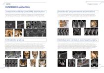
CONE BEAM 3D IMAGING HEAD&NECK applications Temporomandibular joint (TMJ) examination Diagnosis and anatomical assessment of the temporomandibular joint can be performed with high quality 3D images generated by 5G XL. Sagittal and coronal slices provide optimal imaging of the joint zone to identify any pathologies and to assess the difference between condyle height and the mandibular branch. Orthodontic analysis The ideal application for tomographic, panoramic and cephalometric images acquired with 5G XL is in examinations for aesthetic and orthodontic purposes and for the treatment of severe...
Open the catalog to page 10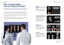
NNT, THE SOFTWARE FOR ALL SPECIALIST NEEDS The versatile and powerful imaging software to perform the examination, process data and share the diagnosis. CONE BEAM 3D IMAGING DENTISTRY: CROSS SECTIONS IN PANORAMIC IMAGES Complete examination of the dental arches in cross sections to check shape, size and status of maxillary and mandibular bones and teeth. NNT is the essential tool to process and manage 2D and 3D images and X-ray videos (CineX). A software that adapts the user interface and offers dedicated analysis functions for the specific needs of radiologists and specialist physicians. Volume...
Open the catalog to page 11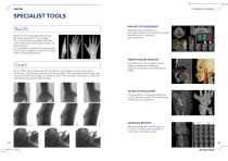
CONE BEAM 3D IMAGING SPECIALIST TOOLS Ray2D With the innovative Ray2D function, 5G XL generates 2D X-ray images (18 x 19 cm) that are perfect for both preliminary and post-surgery follow up examinations. It is possible to repeat the investigation from various angles to select the image with the best perspective. CineX 5G XL offers the exclusive CineX function to investigate moving anatomical structures, such as saliva ducts and joint mobility. This advanced technology uses a sequence of X-ray images to create an 18 x 19 cm format video that can also be exported to standard video format. AIRWAY...
Open the catalog to page 12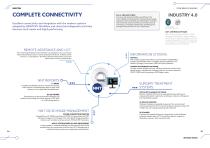
CONE BEAM 3D IMAGING COMPLETE CONNECTIVITY Di.V.A. AND EASY CHECK To ensure maximised workflow smoothness, the Di.V.A. digital virtual assistant provides data and usage statistics to plan workloads and maintenance. The Easy Check tool also ensures continuous remote technical monitoring, to facilitate maintenance scheduling and anticipate the resolution of any critical issues. Excellent connectivity and integration with the modern systems adopted by NEWTOM. Workflow and clinical and diagnostic activities become much easier and highly performing. INDUSTRY 4.0 in according to EN ISO/IEC 17065:2012...
Open the catalog to page 13All NewTom catalogs and technical brochures
 GiANO HR FullView
13 Pages
 DCiS (VET)
7 Pages
 RX DC Wireless (VET)
3 Pages
 RX DC Wired (VET)
3 Pages
 X-PSP (VET)
7 Pages
 5G XL (VET)
11 Pages
 7G (VET)
13 Pages
 7G (MED)
15 Pages
 VGi EVO
17 Pages
 X-PSP (DENT)
7 Pages
 X-VS E (DENT)
5 Pages
 RX DC Wired (DENT)
5 Pages
 DCiS (DENT)
7 Pages
 ViSIOScan WR
7 Pages
 ViSIOScan WL
7 Pages
 VG-One
19 Pages
 GO
GO19 Pages
 Giano HR DC''' (DENT)
21 Pages
- Analysis software
- Visualization software
- Radiology software
- Tablet computer software
- Tablet PC software
- Flat panel detector
- Monitoring software
- Diagnostic software
- Planning software
- Dental software
- Dental radiography system
- Treatment software
- Capture software
- Data management software
- AI-assisted software
- Veterinary X-ray system
- Surgery software
- Artificial intelligence software
- Digital dental radiography system
- Digital veterinary radiography system