Group: Cefla Dental

Catalog excerpts
Cone Beam 3D Imaging
Open the catalog to page 1
First User of Cone Beam in Dental Field QR s.r.l. is the name that stands behind NewTom Cone Beam 3D imaging units and we were the creators of Cone Beam technology for the dental field. NewTom 9000 (also known as Maxiscan) was the very first Cone Beam equipment in the world, which was installed in 1996. It is the forefather of NewTom product line and, in general, of the entire X-Ray units based on Cone Beam technology. QR’s 20 plus years of experience and success in research, development, manufacturing and distribution of NewTom products affirms our commitment to excellence and quality. QR...
Open the catalog to page 2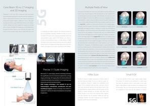
Cone Beam 3D vs. CT Imaging and 2D Imaging Multiple Fields of View The scanner’s FOV determines how much of the patient’s anatomy will be visua- Traditional CT (CAT scan) uses a narrow fan beam that rotates lized. If using a flat panel detector (FPD), the dimensions of their cylindrical FOV can be described as Diameter by Height (DxH). Nowadays, the need to scan dif- around the patient acquiring thin axial slices with each revolution. In ferent R.O.I. (Region Of Interest) with different dimensions is regulated by interna- order to create a section of anatomy, many rotations must be done....
Open the catalog to page 3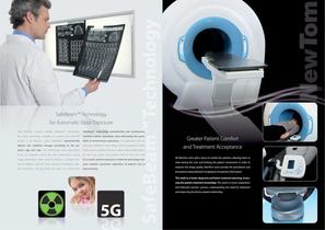
SafeBeam™ Technology for Automatic Dose Exposure Only NewTom systems employ SafeBeam™ technology, SafeBeam™ technology automatically and continuously the safest technology available for patient and staff. Fe- monitors system operations, thus eliminating the possi- atured in all NewTom units, SafeBeam™ automatically bility of unnecessary exposures. In conjunction with our adjusts the radiation dosage according to the pa- patented SafeBeam™ technology, when compared to other tient’s age and size. This technology uses intermittent CB3D systems, NewTom 5G has a wider range of adjustments bursts...
Open the catalog to page 4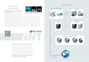
Network Set-Up NewTom NNT Analysis Software NewTom Standard Supply NewTom NNT analysis software is the perfect solution for Cone Beam 3D imaging. NNT allows the creation of different kinds of 2D and 3D images in a 16 bit grey-scale and it takes only few seconds to evaluate the data taken during the scan. The software is totally designed by NewTom engineers and, thanks to the various application modes specifically design for can be delivered digitally (burnt to a CD or DVD), on paper, different fields of use, it fulfills all the requirements and needs film or pdf. The software is available...
Open the catalog to page 5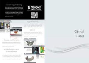
NewTom Implant Planning NewTom Implant Planning is a software package that allows the creation of 3D implant simulation on any PC. It can simulate the implant placement on 2D and 3D models, identify the mandibular canal, draw panoramics and cross sections of the bone model. It also shows the 3D bone model and calculates the bone density. NewTom Implant Planning is used to plan prosthesis implant surgery in a faster, safer and more efficient way. It also allows the ability to export in .stl format. Supported Formats NewTom Implant Planning reads axial slices saved in DICOM 3.0 or in NNT...
Open the catalog to page 6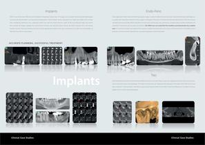
CB3D is one of the most effective tools available for analyzing implant sites. 3D images can accurately identify possible pathologies These application fields need extremely high quality images in order to define the tooth structure, determine the exact pathology and and structural abnormalities. Cross sectional and panoramic views facilitate various calculations as: height and width of the implant accurately plan the perfect treatment. Only a proper investigation of the area of interest will make the dentist aware of the less invasive sites, mandibular edentulous site, a potential implant...
Open the catalog to page 7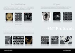
Oral and Maxillofacial Surgery A 3D post-operative scan can show the exact individual anatomy and define the anatomical structures, bringing the patient Due to the multiple FOV and the high level of accuracy in the images, NewTom 5G shows clearly all the airways, structures of the ears, to a better acceptance of the treatment. NewTom scans are useful also for maxillofacial post-surgery imaging, due to TMJ and sinuses using always the proper radiological parameters in order to avoid unnecessary radiation to the patient. The operator reduced image scatter and lower radiation. High Resolution...
Open the catalog to page 8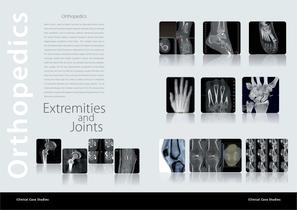
Orthopedics Bone X-ray is used to detect fractures or dislocated joints, ensure that a fracture has been properly aligned, evaluate injury or damage from conditions such as infections, arthritis, abnormal bone growths, locate foreign objects, evaluate changes in bones and detect degenerative conditions of the bone. The multiple views due to the 3D dataset allow specialists to assess the degree of pathological displacement of any fractures or dislocations. Foot X-ray requires an AP view for better viewing the medical aspect of the foot (i.e. talus, navicular, medial and middle cuneiform...
Open the catalog to page 9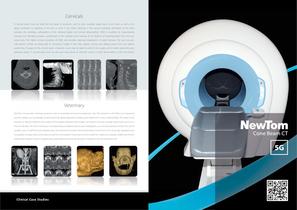
Cervicals A cervical spine X-ray can help find the cause of symptoms such as neck, shoulder, upper back, or arm pain, as well as tingling, numbness, or weakness in the arm or hand. It can detect fractures in the cervical vertebrae, dislocation of the joints between the vertebrae, subluxations of the vertebral bodies and cervical abnormalities. CB3D is excellent for characterizing fractures and identifying osseous compromise of the vertebral canal because of the absence of superimposition from the transverse view. The higher contrast resolution of CB3D also provides improved visualization of...
Open the catalog to page 10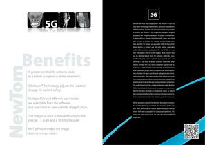
NewTom 5G, from the company that was the first to use the Cone Beam technology in dental field, represents the newest in CB3D technology. NewTom 5G takes an image at every degree of rotation, 360° rotation = 360 images, increasing the range of possibilities for image manipulation. It couples a revolutionary flat panel X-ray detector technology with a very small focal spot (0.3mm), to produce the clearest, sharpest images possible. NewTom 5G features an adjustable Field Of View, which allows doctors to irradiate just the right volume, depending on the different clinical applications. The...
Open the catalog to page 11All NewTom catalogs and technical brochures
-
 NewTom 5G XL
14 Pages
-
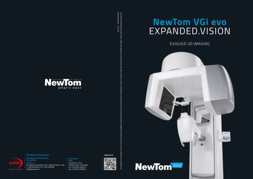 VGi evo
16 Pages
-
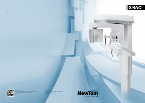 GiANO
GiANO20 Pages
-
 GO
GO18 Pages
-
 7G
7G12 Pages
-
 New Tom X-PSP
12 Pages
-
 GiANO HR
20 Pages
Archived catalogs
-
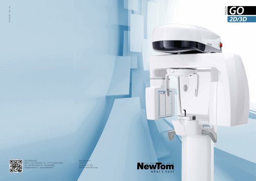 GO 2D/3D
14 Pages
-
 5G
5G10 Pages
-
 VGi
VGi10 Pages
-
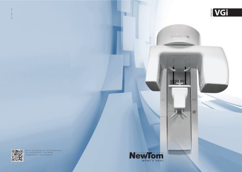 NEWTOM VGi
10 Pages
-
 NewTom GiANO
13 Pages
-
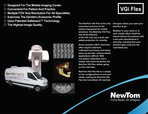 Newtom VGi Flex
2 Pages
