RX DC Wired (VET)
1 /3Pages

RX DC Wired (VET)
1 /3Pages
Catalog excerpts
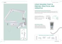
CONE BEAM 3D IMAGING X-RAY IMAGING THAT IS PRECISE, PRACTICAL AND VERSATILE HIGHER PERFORMANCE AND MAXIMUM ERGONOMICS Thanks to the protractor with graduated scale, arm and tube head positioning is stable, effective and just right for the task. Featuring an integrated self-balancing device, allowing them to be pointed in 6 directions, the arms are available in the following lengths: 40, 60 and 90 cm, making installation as simple as possible. OPTIMAL DEFINITION SUB-OPTIMAL DEFINITION FOCAL SPOT FOCAL SPOT Increased X-ray parallelism and an incorporated collimator allow the RX DC VET to achieve...
Open the catalog to page 2
Constant potential, microprocessor-controlled 145 ÷ 230 KHz with self-adjustment (typically 175 KHz) Focal spot Total filtration Anode current Voltage at X-ray tube Exposure times Source-skin distance Irradiated field Ø 60 mm and Ø 55 mm (with round cone) Additional collimators (optional) 35 x 45 mm (with rectangular cone for size 2 sensors) 31 x 41 mm and 22 x 35 mm, for size 1 and size 0 sensors Power supply Duty Cycle Continuous operation with self-adjustment up to 1s/90s total Arms (for Standard version only) Standard (wall mounted) or Mobile (on portable cart) Working frequency (*) values...
Open the catalog to page 3All NewTom catalogs and technical brochures
 GiANO HR FullView
13 Pages
 DCiS (VET)
7 Pages
 RX DC Wireless (VET)
3 Pages
 X-PSP (VET)
7 Pages
 5G XL (VET)
11 Pages
 7G (VET)
13 Pages
 5G XL (MED)
15 Pages
 7G (MED)
15 Pages
 VGi EVO
17 Pages
 X-PSP (DENT)
7 Pages
 X-VS E (DENT)
5 Pages
 RX DC Wired (DENT)
5 Pages
 DCiS (DENT)
7 Pages
 ViSIOScan WR
7 Pages
 ViSIOScan WL
7 Pages
 VG-One
19 Pages
 GO
GO19 Pages
 Giano HR DC''' (DENT)
21 Pages
- Analysis software
- Visualization software
- Radiology software
- Tablet computer software
- Tablet PC software
- Flat panel detector
- Monitoring software
- Diagnostic software
- Planning software
- Dental software
- Dental radiography system
- Treatment software
- Capture software
- Data management software
- AI-assisted software
- Veterinary X-ray system
- Artificial intelligence software
- Surgery software
- Digital dental radiography system
- Digital veterinary radiography system