Group: Cefla Dental

Catalog excerpts
CEFLA s.c. Via Selice Provinciale 23/a • 40026 Imola • Italy t. +39 045 8202727 • 045 583500 info@newtom.it newtom.it Stabilimento Plant According to the standards in force, in extra-EU areas the availability and specifications of some products and/or characteristics may vary. Please contact your local distributor for further information. Pictures are for illustration purpose only. BU Medical Equipment Sede legale ed amministrativa Headquarters NewTom VGi evo EXPANDED.VISION EVOLVED 3D IM
Open the catalog to page 1
FROM NEWTOM’S RESEARCH AND INNOVATION, THE COMPLETE MAXILLOFACIAL/ENT CBCT. Detailed viewing accuracy, superior scanning technology. Multiple FOVs up to 24 x 19 cm for complete Head&Neck 3D diagnostics and 2D examinations in a single scan. The CineX function offers a dynamic view of moving structures. The dose radiated to the patient can be considerably reduced with the ECO Scan mode.
Open the catalog to page 2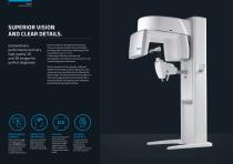
SUPERIOR VISION AND CLEAR DETAILS. Extraordinary performance and very high quality 2D and 3D images for perfect diagnoses. NewTom’s research and experience has produced VGi evo, the versatile and efficient device that offers technology, safety, comfort and a broad range of FOVs for acquisitions up to 24 x 19 cm. A wide range of volumetric, panoramic and teleradiographic examinations as well as dynamic X-rays for perfect diagnoses in all situations. With the exclusive Eco Scan acquisition modes and SafeBeamTM technology, excellent image quality can be ensured with very low radiated doses to...
Open the catalog to page 3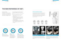
THE NEW REFERENCE OF CBCT. Outstanding definition and quality with the revolutionary NewTom image chain. Complete 360° rotation to rapidly acquire superior quality cylindrical volume with advanced kinematic technology (patented). The technologically advanced elements that form the innovative image chain of VGi evo carry the performance of CBCT devices to a new extraordinary level: • the ultimate large sensor allows to examine a volume up to 24 x 19 cm with increased signal-noise ratio; • the generator with rotating anode and 0.3 mm focal spot allows to obtain very high definition images to...
Open the catalog to page 4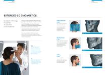
EXTENDED 3D DIAGNOSTICS. Complete FOV range for perfect 3D volumes in all situations. VGi evo is a versatile and effective device with several examination modes dedicated to an extensive selection of clinical applications. The choice of field of view determines the expanse of the anatomical district analysed. VGi evo complies with international standards based on the “ALARA” (As Low As Reasonably Achievable) principle, whose purpose is to reduce the dose absorbed by the patient by selecting the most suitable FOV for the anatomical region of interest. The exclusive Boosted and Enhanced modes...
Open the catalog to page 5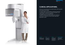
CLINICAL APPLICATIONS. VGi evo is a powerful and versatile device that expands the clinical use of CBCT. Its wide range of examinations meets all needs of maxillofacial, otorhinolaryngology, dentistry and orthopaedic-cervical procedures. The NNT software provides dedicated interfaces and tools to make the most of every specialist physician’s work. CERVICAL REGION FOVs up to 24 x 19 cm for a complete view of the entire maxillofacial area. Very high definition examinations of the internal ear and complete airways. Complete high quality dental applications for implantology, orthodontic...
Open the catalog to page 6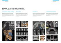
DENTAL CLINICAL APPLICATIONS. ORAL AND MAXILLOFACIAL SURGERY Highly precise details for oral and maxillofacial surgery applications, such as the presence of teeth or fractures, bone density and height, form and inclination of the root. The presence of metal elements does not impact image quality; instead, the small quantity of radiation reduces the scattering effect to a minimum and, therefore, anatomical structures are clearly displayed. Cone Beam technology is ideally applied in orthodontic applications for aesthetic purposes and for the treatment of severe diseases. In fact, 3D images...
Open the catalog to page 7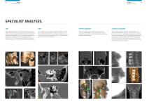
UPPER AIRWAYS CERVICAL REGION High-quality 3D images in the anatomical representation of both the TMJ and the cervical region. Sagittal and coronal slices provide optimal imaging of the joint zone and are essential to identify any pathologies. Panoramic images provide important orthodontic information for initial screening, such as the difference between the height of the condyle and that of the mandibular ramus, or information on other dental pathologies. With a single scan, VGi evo generates HiRes images of airways, both temporomandibular joints, maxillary and nasal sinuses. The clear and...
Open the catalog to page 8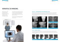
VERSATILE 2D IMAGING. Panoramic and cephalometric examinations for a precise and complete view. The innovative VGi evo technology includes a low dose CBCT scan that has been specially developed for combined use with the patented Sharp 2D function, which generates a complete set of 2D images for diagnostic screening and post-surgery follow-up examinations. Sharp 2D – TELERADIOGRAPHIC AND PANORAMIC SCANS Exclusive function to create a dataset of images from Panoramic and Teleradiographic (AP, PA and LL) scans in a single examination. Compared to the panoramic scan-like coronal reconstructions...
Open the catalog to page 9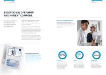
EXCEPTIONAL OPERATOR AND PATIENT COMFORT. Functional features and design that facilitate relations and diagnosis. VGi evo offers excellent ergonomics and stability during scans. The patented head support unit offers 7 contact points for rapid access and natural positioning of the patient. Three laser lines precisely indicate the references of the area of interest. The mirror placed opposite the chin rest, and acquisition of two low dose scout (latero-lateral and antero-posterior) images provide a complete view of the patient, and allow to check correct position and perfect alignment....
Open the catalog to page 10
SOFTWARE NEWTOM.
Open the catalog to page 11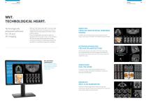
NNT. TECHNOLOGICAL HEART. Technologically advanced software for 2D and 3D imaging. With just a few simple steps NNT can process data acquired during the scan to create a vast array of images, which provide detailed information about patient anatomy. They can subsequently be saved in a report or distributed with the Viewer version of the software. NNT also provides different application modes specifically intended for implantology, endodontics, periodontics, maxillofacial surgery and radiology. DENTISTRY: CROSS SECTIONS IN DENTAL PANORAMIC IMAGING Complete view of the dental arches in cross...
Open the catalog to page 12All NewTom catalogs and technical brochures
-
 NewTom 5G XL
14 Pages
-
 GiANO
GiANO20 Pages
-
 GO
GO18 Pages
-
 7G
7G12 Pages
-
 New Tom X-PSP
12 Pages
-
 GiANO HR
20 Pages
Archived catalogs
-
 GO 2D/3D
14 Pages
-
 5G
5G10 Pages
-
 VGi
VGi10 Pages
-
 NEWTOM VGi
10 Pages
-
 NewTom GiANO
13 Pages
-
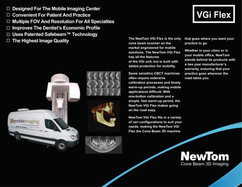 Newtom VGi Flex
2 Pages
-
 NEWTOM 5G
13 Pages
