RS-1 Glauvas
1 /12Pages
RS-1 Glauvas
1 /12Pages
Catalog excerpts
RS-1 Glauvas Specifications OCT scanning Principle Optical resolution Scan width Scan depth OCT light source Scan speed Image averaging Fundus surface imaging Principle Angle of view Internal fixation lamp color External fixation lamp color Auto alignment Minimum pupil diameter Diopter correction range Working distance Software analysis Retina Normative database Axial length Age Scan pattern Long axial length normative database*2 Axial length Age Scan pattern PC networking Power supply Power consumption Dimensions/mass Optional accessories Optical Coherence Tomography Spectral domain OCT Z: 7 µm, X-Y: 20 µm Line: Up to 16.5 mm Map: Up to 15.0 x 12.0 mm 4.2 mm SLD, 880 nm Up to 250,000 A-scans/s Up to 120 images SLO 53.3°(X) x 53.3°(Y) Green Red X-Y-Z directions ø2.5 mm -20 to +20 D (VD=0 mm) Fundus OCT mode: 24.9 mm / Anterior*1: 20.0 mm Segmentation of 7 retinal layers Scan width correction Full retina thickness map GCC thickness map RNFL thickness map Percentile map (RNFL, GCC+RNFL) Structural normality map Thickness map Disc analysis Follow-up analysis Corneal thickness measurement Corneal thickness map Angle measurement Less than 26 mm 20 years to under 80 years Macula map, disc map, retina map 26 mm to less than 29 mm 20 years to under 60 years Macula map, disc map, retina map Available 100 to 240 V AC, 50/60 Hz Device main body 220 VA 332 (W) x 526 (D) x 586 (H) mm / 30.6 kg 13.0 (W) x 20.7 (D) x 23.1 (H)" / 67.5 lbs. B-scan denoising software, OCT-A dongle, computer, computer monitor, isolation transformer, pole-type computer cabinet (slim), anterior segment OCT image capture kit, motorized optical table, HDD *1 Anterior segment OCT image capture kit is optional. *2 Data was collected from a sample of Asian patients. Image courtesy of Lee Shu Yen, MD, Singapore Kelvin Teo Yi Chong, MD, Singapore Retina Foundation & Eye Research Center, India San Giuseppe Hospital - IRCCS MultiMedica, Italy Vista System Center, Italy More clinical information available online at the NIDEK Education page For more clinical information, please visit the Education page on the NIDEK website. This site allows access to case reports, journal articles, and video presentations. Product/model name: Optical Coherence Tomography RS-1 Brochure and listed features of the device are intended for non-US practitioners. The availability of products differs from country to country depending on the status of approval. Specifications may vary depending on circumstances in each country. Specifications and design are subject to change without notice. HEAD OFFICE [Manufacturer] Distributor in your country 34-14 Maehama, Hiroishi-cho, Gamagori, Aichi 443-0038, JAPAN Phone: +81-533-67-8895 Group Website www.nidek.com Product Information www.nidek-intl.com/product/ Please contact our distributor for more information. www.nidek-intl.com/dist/
Open the catalog to page 1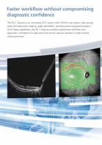
Faster workflow without compromising diagnostic confidence The RS-1 Glauvas is an innovative OCT system with 250kHz scan speed, high-quality wide and deep area imaging, great operability, and deep learning-based analytics. With these capabilities, the RS-1 Glauvas provides streamlined workflow and diagnostic confidence for glaucoma and retinal vascular diseases in high-volume clinical practices. 250,000 A-scans/s high-speed imaging Wide, deep, highresolution imaging Effortless operation and interpretation Advanced analytics
Open the catalog to page 2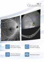
Faster workflow without compromising diagnostic confidence The RS-1 Glauvas is an innovative OCT system with 250kHz scan speed, high-quality wide and deep area imaging, great operability, and deep learning-based analytics. With these capabilities, the RS-1 Glauvas provides streamlined workflow and diagnostic confidence for glaucoma and retinal vascular diseases in high-volume clinical practices. 250,000 A-scans/s high-speed imaging Wide, deep, highresolution imaging Effortless operation and interpretation Advanced analytics
Open the catalog to page 3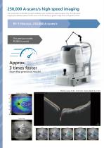
250,000 A-scans/s high-speed imaging Macula line 16.5 mm / Scan depth 4.2 mm The incorporation of 250,000 A-scans/s accelerates your workflow by reducing capture time. The high-speed imaging also addresses patient fixation errors thus contributing to greater image clarity and patient comfort. The previous model: 85,000 A-scans/s Approx. 3 times faster than the previous model Retina map 15.0 x 12.0 mm / Scan depth 4.2 mm Wide, deep, highresolution imaging With RS-1 Glauvas, a single B-scan image clearly presents the area from the optic nerve head to the temporal vascular arcade, and the 4.2 mm...
Open the catalog to page 4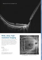
250,000 A-scans/s high-speed imaging Macula line 16.5 mm / Scan depth 4.2 mm The incorporation of 250,000 A-scans/s accelerates your workflow by reducing capture time. The high-speed imaging also addresses patient fixation errors thus contributing to greater image clarity and patient comfort. The previous model: 85,000 A-scans/s Approx. 3 times faster than the previous model Retina map 15.0 x 12.0 mm / Scan depth 4.2 mm Wide, deep, highresolution imaging With RS-1 Glauvas, a single B-scan image clearly presents the area from the optic nerve head to the temporal vascular arcade, and the 4.2 mm...
Open the catalog to page 5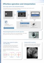
Effortless operation and interpretation Easy image capture with automated functions Intuitive user interface The auto alignment and auto switch functions allow anyone to effortlessly capture images. Operators need to only adjust the chinrest height and click Optimize and Capture. The OCT Viewer software allows quick access to images and analytics. The display can be viewed by day, scan pattern, and analysis mode within a single screen - allowing faster review of imaging and analytics. Auto alignment Right eye / both eyes / left eye Analysis mode Summary, glaucoma, and macular analysis modes are...
Open the catalog to page 6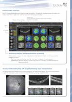
Effortless operation and interpretation Easy image capture with automated functions Intuitive user interface The auto alignment and auto switch functions allow anyone to effortlessly capture images. Operators need to only adjust the chinrest height and click Optimize and Capture. The OCT Viewer software allows quick access to images and analytics. The display can be viewed by day, scan pattern, and analysis mode within a single screen - allowing faster review of imaging and analytics. Auto alignment Right eye / both eyes / left eye Analysis mode Summary, glaucoma, and macular analysis modes are...
Open the catalog to page 7All NIDEK catalogs and technical brochures
 Edging Station LE-800
6 Pages

Archived catalogs
 SL-1800
2 Pages
 Nex-Acri
4 Pages
 OPD-Scan III
6 Pages
 Auto Lensmeter LM-500
4 Pages
 ConfoScan4 CS4
2 Pages
 EC-5000CXlll Specifications
16 Pages
 Satellite Tracer Lt 910
2 Pages
 System Chart SC-1600
4 Pages
 System edger SE-9090
12 Pages
 Echoscan US-4000/500
4 Pages


- Ultrasound system
- Syringe
- B/W ultrasound system
- Tabletop laser
- Nd:YAG laser
- On-platform ultrasound system
- Fixed ophthalmic examination
- Touchscreen ultrasound system
- Hand-held ophthalmic examination instrument
- Slit lamp
- Nanosecond laser
- Table slit lamp
- Ophthalmoscope
- Ophthalmic laser
- Automatic optical lens processing system
- Tonometer
- Retinal camera
- Refractometer ophthalmic examination
- Automatic refractometer
- Keratometer ophthalmic examination