Scanning Laser Ophthalmoscope Mirante SLO/OCT Mirante SLO
1 /16Pages
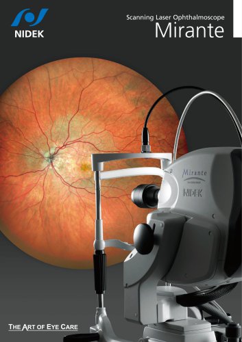
Scanning Laser Ophthalmoscope Mirante SLO/OCT Mirante SLO
1 /16Pages
Catalog excerpts

Mirante Specifications Anterior*4 Common specification Diopter correction range Internal fixation lamp External fixation lamp Tilt Swing PC networking Power supply Power consumption Dimensions/mass*5 Optional accessories Ultra wide field: 163°* Angle of incidence Ultra wide field: 110° Angle of incidence Standard: 60° * Measured from the center of the eye X: 3 to 16.5 mm, Y: 3 to 13.2 mm, Z: 2.1 mm X: 2 to 8 mm, Z: 2.1 mm SLD, 880 nm Up to 85,000 A-scans/s Up to 120 images 9 x 9 mm (macula), 6 x 6 mm (disc) ø2.5 mm Standard: 19 mm / Anterior*4: 15.4 mm Segmentation of 6+1 retinal layers, macular thickness map, RNFL thickness map, [NFL+GCL+IPL] analysis, optic nerve analysis Corneal thickness measurement, corneal thickness map, angle measurement -15 to +15 D Red (670 nm) / blue (488 nm) White ±10° ±20° Available 100 to 240 V AC, 50/60 Hz Device main body 150 VA 345 (W) x 548 (D) x 527 to 557 (H) mm / 23 kg (SLO/OCT model) 22 kg (SLO model) 13.6 (W) x 21.6 (D) x 20.7 to 21.9 (H)" / 51 lbs. (SLO/OCT model) 49 lbs. (SLO model) Wide-field adapter, motorized optical table, PC rack, isolation transformer, external fixation lamp (multi-joint), anterior segment OCT adapter*3, AngioScan (OCT-Angiography)*3, long axial length normative database*3, B-Scan Denoising Software*3, FA/ICG dongle*6, FA dongle*6 *1 Ultra wide field imaging is available with the optional wide-field adapter. *2 Optional for the SLO model. *3 Available for the SLO/OCT model. *4 Anterior segment OCT adapter is optional. *5 Only for image capturing unit. *6 Available for the SLO model. Video size*2 Minimum pupil diameter Working distance OCT*3 Principal Optical resolution Scan range Retina Anterior*4 OCT light source Scan speed Image averaging Normative database Minimum pupil diameter Working distance Software analysis Retina Scanning Laser Ophthalmoscope Central angle of view Confocal scanning Standard: Diagonal angle of view 89° Ultra wide field*1: ø163° 488, 532, 670, 790 nm 4,096 x 4,096, 2,048 x 2,048, 1,536 x 1,536, 1,024 x 1,024, 768 x 768, 512 x 512 (pixel x pixel) 1,024 x 1,024, 768 x 768, 512 x 512 (pixel x pixel) ø3.3 mm Standard: 19 mm / Ultra wide field*1: 9 mm SLO Principal Angle of view (Measured from the center of the eye) Light source Still image size Images courtesy of Luigi Sacco Hospital, University of Milan, Italy Asia Eye Centre, Singapore Doheny Eye Center, UCLA, USA Retina Foundation & Eye Research Center, India Kagoshima University Hospital, Japan Exilaser Clinic, Peru Chiba University Hospital, Japan Tohoku University, Japan Careggi University Hospital, University of Florence, Italy More clinical information available online at the NIDEK Education page For more clinical information, please visit the Education page on the NIDEK website. This site allows access to case reports, journal articles, and video presentations. Product/model name: Scanning Laser Ophthalmoscope Mirante Brochure and listed features of the device are intended for non-US practitioners. Specifications may vary depending on circumstances in each country. Specifications and design are subject to change without notice. HEAD OFFICE (International Div.) TOKYO OFFICE (International Div.) 34-14 Maehama, Hiroishi-cho, Gamagori, Aichi 443-0038, JAPAN TEL: +81-533-67-8895 URL: www.nidek.com 3F Sumitomo Fudosan Hongo Bldg., 3-22-5 Hongo, Bunkyo-ku, Tokyo 113-0033, JAPAN TEL: +81-3-5844-2641 URL: www.nidek.com 2040 Corporate Court, San Jose, CA 95131, U.S.A. TEL: +1-408-468-6400 +1-800-223-9044 (US Only) URL: usa.nidek.com Ecoparc, 9 rue Benjamin Franklin, 94370 Sucy En Brie, FRANCE TEL: +33-1-49 80 97 97 URL: www.nidek.fr Via dell’Artigianato, 6/A, 35020 Albignasego (Padova), ITALY TEL: +39 049 8629200 / 8626399 URL: www.nidektechnologies.it Rm3205,Shanghai Multi Media Park, No.1027 Chang Ning Rd, Chang Ning District, Shanghai, CHINA 200050 TEL: +86 021-5212-7942 URL: www.nidek-china.cn 51 Changi Business Park Central 2, #06-14, The Signature 486066, SINGAPORE TEL: +65 6588 0389 URL: www.nidek.sg
Open the catalog to page 1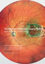
Retro mode The Ultimate Multimodal Imaging Platform State-of-the-art SLO/OCT Combo Ultra Wide Field x Ultra HD image A stellar combination of 163° ultra wide field x ultra 4K HD incorporated in the Mirante achieves a wider, enhanced view of the retinal structure and vasculature with unparalleled clarity. (Ultra wide field image is available with the optional wide-field adapter.) The FlexTrack technology improves imaging quality. *1 Available for the SLO/OCT model. Optional for the SLO model. *2 Available for the SLO/OCT model. *3 Optional for the SLO/OCT model.
Open the catalog to page 2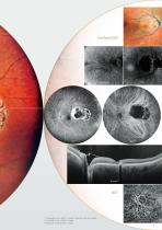
Retro mode The Ultimate Multimodal Imaging Platform State-of-the-art SLO/OCT Combo Ultra Wide Field x Ultra HD image A stellar combination of 163° ultra wide field x ultra 4K HD incorporated in the Mirante achieves a wider, enhanced view of the retinal structure and vasculature with unparalleled clarity. (Ultra wide field image is available with the optional wide-field adapter.) The FlexTrack technology improves imaging quality. *1 Available for the SLO/OCT model. Optional for the SLO model. *2 Available for the SLO/OCT model. *3 Optional for the SLO/OCT model.
Open the catalog to page 3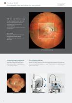
Mirante SLO/OCT Mirante SLO Unsurpassed color and clarity for every detail 163° ultra wide field color image The clear image of the entire 163° field The FlexTrack algorithm corrects image of view enables detailed evaluation of distortion due to unstable fixation and pathologies from the fovea to the enhances averaging quality. extreme periphery. (Ultra wide field imaging is available with the optional wide-field adapter.) Refine mode As required, capturing two images with Ultra 4K HD and averaging function for unparalleled clarity slightly different fixation reduces reflection, producing a clear...
Open the catalog to page 4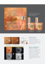
Mirante SLO/OCT Mirante SLO Unsurpassed color and clarity for every detail 163° ultra wide field color image The clear image of the entire 163° field The FlexTrack algorithm corrects image of view enables detailed evaluation of distortion due to unstable fixation and pathologies from the fovea to the enhances averaging quality. extreme periphery. (Ultra wide field imaging is available with the optional wide-field adapter.) Refine mode As required, capturing two images with Ultra 4K HD and averaging function for unparalleled clarity slightly different fixation reduces reflection, producing a clear...
Open the catalog to page 5All NIDEK catalogs and technical brochures
 Edging Station LE-800
6 Pages
 RS-1 Glauvas
12 Pages

Archived catalogs
 SL-1800
2 Pages
 Nex-Acri
4 Pages
 OPD-Scan III
6 Pages
 Auto Lensmeter LM-500
4 Pages
 ConfoScan4 CS4
2 Pages
 EC-5000CXlll Specifications
16 Pages
 Satellite Tracer Lt 910
2 Pages
 System Chart SC-1600
4 Pages
 System edger SE-9090
12 Pages
 Echoscan US-4000/500
4 Pages


- Ultrasound system
- Syringe
- B/W ultrasound system
- Tabletop laser
- Nd:YAG laser
- On-platform ultrasound system
- Fixed ophthalmic examination
- Touchscreen ultrasound system
- Hand-held ophthalmic examination instrument
- Slit lamp
- Nanosecond laser
- Table slit lamp
- Ophthalmic laser
- Automatic optical lens processing system
- Tonometer
- Retinal camera
- Refractometer ophthalmic examination
- Automatic refractometer
- Keratometer ophthalmic examination