- Catalogs
- Nikon Instruments
- A1 MP+ / A1R MP+
- Products
- Catalogs
- News & Trends
- Exhibitions
A1 MP+ / A1R MP+
1 /20Pages

A1 MP+ / A1R MP+
1 /20Pages
Catalog excerpts
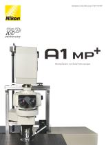
Multiphoton Confocal Microscope A1 MP+/A1R MP+ i m MP+ Multiphoton Confocal Microscope
Open the catalog to page 1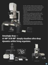
The A1 MP+ and A1R MP+ multiphoton confocal microscopes provide faster and sharper imaging from deeper within living organisms, extending the boundaries of traditional research techniques in biological sciences. • Deep specimen imaging with ultrasensitive GaAsP non-descanned detectors (GaAsP NDDs) located close to the back aperture of the objective lens. In particular, 1300 nm-compatible episcopic GaAsP NDDs for upright microscopes allow deep in vivo imaging of mouse brains up to 1.4 mm. •A 1300 nm-compatible A1R MP+ scan head which is capable of simultaneous excitation with two wavelenghts is...
Open the catalog to page 3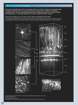
Ultra-deep imaging of living specimens Ultrasensitive GaAsP NDDs allow clear in vivo imaging in deeper areas than ever before and are powerful enough to analyze fast dynamics, such as the activation of neurons, in living specimens. In addition to the 1080 nmcompatible model, a 1300 nm-compatible model GaAsP NDD for upright microscopes is also available, enabling deep imaging of up to 1.4 mm when combined with a 1300 nm-compatible A1R MP+ scan head. Deep brain imaging in in vivo mouse with 1300 nm-compatible GaAsP NDD In vivo imaging of an anesthetized YFP-H mouse (4-week-old) via open skull method....
Open the catalog to page 4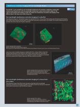
Simultaneous excitation imaging with two-wavelength IR laser The A1R MP+ is now available for two-wavelength simultaneous IR excitation. Combining a system with a femtosecond IR pulse laser with two wavelength simultaneous output (main tunable output of 700 – 1300 nm and auxiliary fixed output of 1040 nm) enables the simultaneous excitation and imaging of two different dyes in the deep areas within living cells. Two wavelength simultaneous excitation imaging of a zebrafish Three dimensional images of 1 dpf zebrafish transgenic line, Tg[h2afv:GFP; EF1α : mCherry-zGem]. After breeding under Phenyltiourea...
Open the catalog to page 5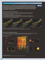
Channel unmixing Fast multiphoton imaging, powerful enough for in vivo imaging The Nikon resonant scanner is equipped with A1R MP+ and capable of high-speed 420 fps (512 x 32 pixel) imaging. Unique to this design is a resonant scan mirror capable of imaging full fields of view at much higher speeds than traditional galvano scanners. Nikon's optical pixel clock system, which monitors the position of the resonant mirror in real time, adjusts the pixel clock to ensure more stable, geometrically correct and more evenly illuminated imaging, even at high speeds. This enables the successful visualization...
Open the catalog to page 6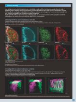
Channel unmixing With multiphoton excitation, fluorophores have a considerably broader profile of the absorption spectra than with single photon excitation. Therefore, simultaneous excitation of multiple fluorophores with a single excitation wavelength is possible. Additionally, the wavelength of a pulsed laser for multiphoton excitation can be changed and the user can select a suitable and well-balanced wavelength for the excitation of multiple fluorophores. A1 MP+/A1R MP+ NDD and channel unmixing technology enable the user to clearly isolate multiple fluorophores and obtain information on the...
Open the catalog to page 7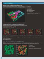
Channel unmixing Multiphoton imaging gallery Four-color imaging of human colon cancer cells in in vivo Three-dimensional volume rendering of implanted subcutaneous tumor of HCT116 expressing Fucci. The cell cycle of tumor cells and the environment (collagen fiber and vessels) are visualized. Upper right, only collagen fiber and vessels are shown. Red: Fucci mkO2/cancer cell Green: Fucci mAG/cancer cell Cyan: SHG/collagen fiber Magenta: Qtracker655/neovascular vessels Objective: CFI Plan Fluor 20xA MI Excitation Wavelength: 940 nm Photographed with the cooperation of Drs. Yoshinori Kagawa and...
Open the catalog to page 8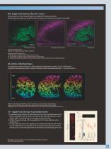
SHG image of the brain surface of a mouse The neocortex of an H-line 5-week-old mouse was studied with the open skull method. The SHG signals from dura mater and EYFP fluorescence signals were simultaneously acquired using the NDD. EYFP fluorescent image SHG image of the dura mater Overlay image Excitation wavelength: 950 nm Objective: CFI75 Apochromat 25xW MP (NA 1.10 WD 2.0) Photographed with the cooperation of: Dr. Takeshi Imamura, Graduate School of Medicine, Ehime University Drs. Yusuke Oshima and Shigenori Nonaka, National Institute for Basic Biology Drs. Terumasa Hibi, Ryoshuke Kawakami...
Open the catalog to page 9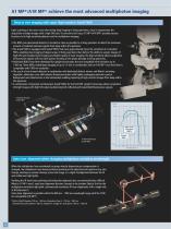
A1 MP+/A1R MP+ achieve the most advanced multiphoton imaging Deep in vivo imaging with super high-sensitive GaAsP NDD Light scattering is the main issue when doing deep imaging in living specimens, since it compromises the acquisition of bright images with a high S/N ratio. To overcome this issue, A1 MP+/A1R MP+ provides various functions in its high sensitivity detector unit for multiphoton imaging: • he NDD (non-descanned detector) is located as close as possible to a living specimen to detect the maximum T amount of scattered emission signals from deep within the specimen. • he GaAsP NDD is...
Open the catalog to page 10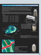
Nikon’s high-NA objectives are ideal for multiphoton imaging High-NA objectives that effectively correct chromatic aberrations over a wide wavelength range from ultraviolet to infrared have been developed. Transmission is increased through the use of Nikon’s exclusive Nano Crystal Coat technology. In particular, the CFI Apochromat 25xW MP/MP1300 objective lenses provide the industry’s highest numerical aperture of 1.10, while still maintaining a 2.0 mm working distance. They also have a collar that corrects spherical aberrations depending on the depth of the specimen, and a 33° manipulator pipette...
Open the catalog to page 11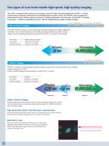
The A1 MP+ is equipped with a galvano (non-resonant) scanner for high-resolution imaging. The A1R MP+ is a hybrid scan head that incorporates both galvano and ultrahigh-speed resonant scanners. The A1R MP+ allows imaging and photoactivation at the ultrafast speeds necessary for revealing cell dynamics and interaction. The A1R MP+ is available in two types - a 1080 nm-compatible type and a 1300 nm-compatible type capable of deeper imaging. The A1 MP+/A1R MP+ galvano scanner enables high-resolution imaging of up to 4096 x 4096 pixels. In addition, with its newly developed scanner driving and sampling...
Open the catalog to page 12All Nikon Instruments catalogs and technical brochures
 Super Resolution Microscopes
28 Pages
 HCA
HCA8 Pages
 DS-Fi3
16 Pages
 A1 HD25 / A1R HD25
15 Pages
 TI-E
TI-E32 Pages
 ECLIPSE TS2
8 Pages
 ECLIPSE TS2R
8 Pages
 N-SIME
6 Pages
 N-SIM N-STORM
24 Pages
 ECLIPSE E100 LED
8 Pages
 ECLIPSE Ti2
24 Pages
 DIGITAL SIGHT SERIES
16 Pages
 Eclipse LV100N-POL Ci-POL
8 Pages
 Eclipse Ti
32 Pages
 C2 Plus
16 Pages
 Biological Microscopes
24 Pages
 Stereomicroscopes
32 Pages
 SMZ18
SMZ1816 Pages
 N-SIM E
4 Pages


Archived catalogs
 A1 MP+ / A1R MP
15 Pages
 ShuttlePix
8 Pages
 Eclipse FN1
12 Pages
 NeoScope Brochure
16 Pages
 ShuttlePix Brochure
5 Pages
 Bio Station IMQ
8 Pages
 MiBrochure
2 Pages
 AZ_C1
AZ_C12 Pages
 High Content Microscopy
2 Pages
 C2+_2CE-SCHH-5
16 Pages
 A1+_A1R+_2CE-SBTH-9
13 Pages
 N-STORM 4.1
13 Pages
 A1_MP
A1_MP11 Pages
 A1+ / A1R+
13 Pages
 NIS-Elements
8 Pages
 Digital Sight Series
9 Pages
 AZ100M
8 Pages
 ShuttlePix P-400R
5 Pages
 Eclipse Ni
15 Pages
 Eclipse Ci-E/ Ci-L
5 Pages
 AZ100
AZ1004 Pages
 Eclipse E200POL
3 Pages
 Eclipse LV100ND POL/DS
2 Pages
 Eclipse E100 LED
4 Pages
 Eclipse E200 POL
3 Pages
 Biological Microscopes
12 Pages
 ECLIPSE LV100N POL 50i/ POL
4 Pages
 Eclipse E100-LED
8 Pages
 A1 MP+ / A1R MP+
11 Pages
 Digital Sight Series Brochure
16 Pages
- Analysis software
- Compound microscope
- Laboratory microscope
- Desktop microscope
- Visualization software
- Tablet computer software
- Tablet PC software
- Control software
- LED microscope
- Laboratory software
- CMOS camera
- Camera with USB port
- LED light source
- Capture software
- Biology microscope
- Training software
- Microscope camera
- HD camera
- Binocular microscope