- Catalogs
- Nikon Instruments
- A1 MP+ / A1R MP
- Products
- Catalogs
- News & Trends
- Exhibitions
A1 MP+ / A1R MP
1 /15Pages

A1 MP+ / A1R MP
1 /15Pages
Catalog excerpts
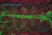
The A1 HD25/A1R HD25 confocal microscope is equipped with an industry-leading field of view of 25mm. There is an ever-increasing demand in life science research for analyses of tissues, organs and live model organisms in order to obtain important biological data. Capturing images of such large samples requires the expansion of areas in which cellular responses can be detected and the reduction of acquisition times. The large field of view of the A1 HD25/A1R HD25 enables image acquisition of large samples in a single scan, extending the boundaries of scientific research.
Open the catalog to page 2
See more than before in confocal resolution With nearly twice the field of view of conventional point scanners, the A1 HD25/A1R HD25 enables users to obtain significantly more data by capturing more of the specimen in each shot.
Open the catalog to page 3
Fewer total images required for large high-resolution image stitching Together with the Ti2-E inverted microscope, the A1 HD25/A1R HD25 is capable of high-quality 25 mm FOV images that capture nearly twice the imaging area of conventional point scanners in each stage position, enabling the acquisition of more spatial information in a single image than ever before. The large FOV reduces both the required number of images for stitching large images and image acquisition time, enabling efficient and high-throughput imaging even with large samples such as live model organisms, tissues and organs....
Open the catalog to page 4
Increase the throughput of your research without compromising image resolution High-speed acquisition of entire wells (with a 4X objective) The combination of a high-speed resonant scanner and large field of view is an ideal platform for high resolution screening assays. It dramatically reduces the time needed to analyze multiple samples and conditions. 8 Large FOV enables measurement of larger areas and high-throughput analysis.
Open the catalog to page 5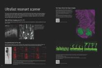
Ultrafast resonant scanner The resonant scanner technologies incorporated in the A1R HD25 produce high-resolution, high-speed imaging at unparalleled levels. The A1R HD25 reduces photobleaching and can acquire the best images for high throughput live cell imaging at high resolutions or multi-dimensional dynamic imaging for applications such as time-lapse and multi-stage position time-lapse experiments. Fast large-volume time-lapse imaging Secretion of Paneth cell granule in response to carbachol was acquired by highspeed 4D live imaging (acquisition of 61 steps of Z-stack images at 1.98 s/ volume...
Open the catalog to page 6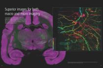
Superior images for both macro and micro imaging Capture large-scale overview images as well as high magnification images with the same instrument. The 25mm FOV of the A1 HD25/A1R HD25 is effective for observation of large samples, while its 1Kx1K high-definition is ideal for the observation of minute structures. Stitched overview image of marmoset brain captured with CFI Plan Apochromat Lambda 10X objective and detailed image of dendritic spines captured with CFI SR HP Plan Apochromat Lambda S 100XC Sil objective
Open the catalog to page 7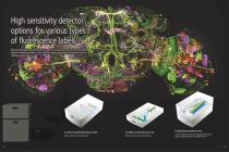
High sensitivity detector options for various types of fluorescence labels Nikon provides highly sensitive detectors for even low intensity specimens. Various wavelength detection methods that support a wide range of imaging applications are available to suit your individual research needs. A1-DUG-2 GaAsP Multi Detector Unit High-sensitivity 4-channel detection A1-DUVB-2 GaAsP Detector Unit Flexible detection with high sensitivity A1-DUS Spectral Detector Unit Accurate separation of closely overlapping fluorescence spectra and the elimination of autofluorescence
Open the catalog to page 8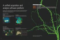
A unified acquisition and analysis software platform Dedicated software modules NIS-Elements C-ER Higher resolution images can be generated with a single click. The software assesses the captured image and automatically determines processing parameters to achieve increased resolution. The unique image processing technology increases image resolution beyond that of a conventional confocal image (resolution can be improved 1.5 times (XY), 1.7 times (Z)). NIS-Elements C, Nikon's unified software platform, provides intuitive workflow for confocal imaging. With graphical programming tools for automating...
Open the catalog to page 9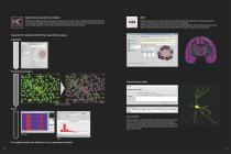
High-Content Acquisition and Analysis With fully-automated acquisition and analysis of a large number of high-content, multidimensional images following an easy stepwise workflow, HCA offers quick experimental setups and an immediate view of measurement data, well by well, during acquisition and via a heat map for trend observation and further analysis. Enables the easy creation of more complex and customized experimental templates, from image acquisition to analysis, without the need for advanced data programming knowledge. JOBS automatically conducts observation processes such as image acquisition,...
Open the catalog to page 10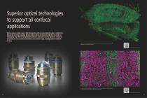
Superior optical technologies to support all confocal applications Nikon provides a broad range of high-NA objectives with unrivaled optical quality to redefine the boundaries of confocal imaging. Options include silicone oil immersion objectives for thick live cell imaging, large-FOV low-magnification objectives and easy-to-use dry objectives. Chromatic aberrations are corrected from ultraviolet to near infrared range, enabling excellent multicolor imaging. Detailed image of deep hippocampus cleared with RapiClear/SunJin Lab and captured with CFI Apochromat LWD Lambda S 20XC WI Scan the QR code...
Open the catalog to page 11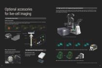
Optional accessories for live-cell imaging For high-speed live cell imaging during photostimulation A new point photostimulation module, available for the Ti2-LAPP modular illumination system, allows the A1 HD25/A1R HD25 to acquire confocal images while simultaneously stimulating the desired area of a sample. TIRF module, DMD module, and epi-fluorescence module are also available for the LAPP system. For long-time observation Perfect Focus System With the Ti2-E inverted microscopes, the Perfect Focus System (PFS) automatic focus maintaining mechanism can be used. It continuously corrects focus...
Open the catalog to page 12
A1ways Evolving Nikon's capacity to respond to customer requirements has resulted in a continuously updated confocal system that exhibits outstanding performance. A1's latest innovation is the addition of a large 25mm field of view, opening new frontiers in research. • Large 25mm field of view • Video rate high-speed imaging H • ybrid scanner for simultaneous photoactivation and imaging • 32-channel spectral imaging D • UVB filterless compact spectral detector H • igh sensitivity GaAsP detector N • IS-Elements C-ER resolution enhancing software C • ombination with Super Resolution Microscopes
Open the catalog to page 13All Nikon Instruments catalogs and technical brochures
 Super Resolution Microscopes
28 Pages
 HCA
HCA8 Pages
 DS-Fi3
16 Pages
 A1 HD25 / A1R HD25
15 Pages
 TI-E
TI-E32 Pages
 ECLIPSE TS2
8 Pages
 ECLIPSE TS2R
8 Pages
 N-SIME
6 Pages
 N-SIM N-STORM
24 Pages
 ECLIPSE E100 LED
8 Pages
 ECLIPSE Ti2
24 Pages
 A1 MP+ / A1R MP+
20 Pages
 DIGITAL SIGHT SERIES
16 Pages
 Eclipse LV100N-POL Ci-POL
8 Pages
 Eclipse Ti
32 Pages
 C2 Plus
16 Pages
 Biological Microscopes
24 Pages
 Stereomicroscopes
32 Pages
 SMZ18
SMZ1816 Pages
 N-SIM E
4 Pages


Archived catalogs
 ShuttlePix
8 Pages
 Eclipse FN1
12 Pages
 NeoScope Brochure
16 Pages
 ShuttlePix Brochure
5 Pages
 Bio Station IMQ
8 Pages
 MiBrochure
2 Pages
 AZ_C1
AZ_C12 Pages
 High Content Microscopy
2 Pages
 C2+_2CE-SCHH-5
16 Pages
 A1+_A1R+_2CE-SBTH-9
13 Pages
 N-STORM 4.1
13 Pages
 A1_MP
A1_MP11 Pages
 A1+ / A1R+
13 Pages
 NIS-Elements
8 Pages
 Digital Sight Series
9 Pages
 AZ100M
8 Pages
 ShuttlePix P-400R
5 Pages
 Eclipse Ni
15 Pages
 Eclipse Ci-E/ Ci-L
5 Pages
 AZ100
AZ1004 Pages
 Eclipse E200POL
3 Pages
 Eclipse LV100ND POL/DS
2 Pages
 Eclipse E100 LED
4 Pages
 Eclipse E200 POL
3 Pages
 Biological Microscopes
12 Pages
 ECLIPSE LV100N POL 50i/ POL
4 Pages
 Eclipse E100-LED
8 Pages
 A1 MP+ / A1R MP+
11 Pages
 Digital Sight Series Brochure
16 Pages
- Compound microscope
- Laboratory microscope
- Desktop microscope
- Visualization software
- Tablet computer software
- Tablet PC software
- Control software
- LED microscope
- Laboratory software
- CMOS camera
- Camera with USB port
- LED light source
- Capture software
- Biology microscope
- Training software
- Microscope camera
- HD camera
- Binocular microscope