

- Products
- Catalogs
- News & Trends
- Exhibitions
A1_MP

A1_MP
- Imaging speed up to 420 fps at 512 x 32 pixels resolution.
- Deep imaging with high-sensitive non-descanned detectors (NDD) and GaAsP NDD model for 1300 nm wavelength.
- Auto laser alignment for correcting IR laser beam shifts.
- Compatibility with upright and inverted microscopes.
- GaAsP NDD enables in vivo imaging up to 1.4 mm deep, beneficial for brain research.
- High clarity imaging of mouse brain structures.
- Allows simultaneous excitation of multiple fluorophores with a single wavelength.
- Supports three-color simultaneous excitation for complex biological studies.
- Resonant scanner for capturing rapid biological processes with minimal photodamage.
- Visualization of intravital microcirculation and blood flow dynamics.
- High-NA objectives with Nano Crystal Coat technology for improved image quality.
- Auto laser alignment for optimal performance with changing wavelengths.
- Ideal for deep multiphoton imaging and physiology research, especially in neuroscience and live tissue studies.
- Applicable in cancer cell imaging and dynamic in vivo studies.
The A1 MP+ series includes features like auto laser alignment for precision, GaAsP NDD for enhanced sensitivity, and hybrid scanning technology for flexible imaging. High-speed data transfer is supported by a fiber-optic system, and optical innovations improve fluorescence efficiency.
Includes galvano and resonant scanners for high-resolution and ultrafast imaging. The hybrid scanning head allows switching between scanners for diverse imaging needs.
Fiber-optic communication supports up to 4 Gbps data transfer, with resonant scanner design preventing overheating.
Features like low-angle incidence dichroic mirrors and hexagonal pinhole design enhance image quality. Dual Integration Signal Processing (DISP) improves signal-to-noise ratio.
Supports high-speed spectral acquisition with real-time unmixing and enhanced diffraction efficiency through the DEES system.
NIS-Elements C software offers intuitive control, including auto laser alignment and Z-intensity control, supporting synchronized imaging with electrophysiology recordings.
Uses high photon density pulsed beams for deep tissue imaging with minimal photo damage, enhanced by pre-chirping systems and non-descanned detectors.
Details on detector types, fluorescence detector wavelengths, filter cubes, and recommended wavelengths for observation. The field of view is a square inscribed in an 18 mm circle, with 4096 gray intensity levels (12 bit).
Supports standard and high-speed acquisition with various scanning modes. The spectral detector range is 400-750 nm.
Compatible with ECLIPSE Ti-E, FN1, and Ni-E microscopes, with optional motorized stages and high-speed positioning systems.
Supports 2D, 3D, and 4D analysis, with applications like FRAP, FLIP, and FRET. Runs on Windows 7 Professional 64-bit.
Compact laser units for multiphoton and confocal microscopy, with specific models and configurations detailed.
Requires specific temperature and humidity conditions, with a dark room or light shield for optimal performance.
Dimensions and weight for components like the scan head, optical units, controllers, and laser units are provided.
Catalog excerpts

Multiphoton Confocal Microscope A1 MP+/A1R MP+ NIKON CORPORATION Fuji Bldg., 2-3, Marunouchi 3-chome, Chiyoda-ku, Tokyo 100-8331, Japan www.nikon.com/ Multiphoton confocal microscope
Open the catalog to page 1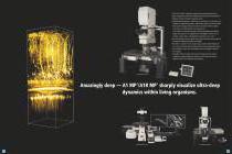
The A1 MP+/A1R MP+ multiphoton confocal microscopes provide faster and sharper imaging from deeper within living organisms, extending the boundaries of traditional research techniques in biological sciences. •Ultrahigh-speed imaging up to 420 frames per second (fps) (512 x 32 pixels) with multiphoton imaging using A1R MP+’ high efficiency optics and resonant scanner. • Deep specimen imaging with high-sensitive non-descanned detectors (NDD) located close to the back aperture of the objective lens. A new model of an ultrasensitive gallium arsenide phosphide (GaAsP) NDD for upright microscopes that...
Open the catalog to page 2
Ultra-deep imaging of living specimens The ultrasensitive GaAsP NDD allows clear in vivo imaging in deeper areas than ever before and is powerful enough to analyze the mechanisms, such as brain neurons, of living specimens. In addition to a model compatible with a wavelength of 1080 nm, there is a new one for upright microscopes that is compatible with a wavelength of 1300 nm. This new NDD enables deep imaging up to 1.4 mm in combination with a newly developed scanning head A1R MP+ that is compatible with a wavelength of 1300 nm. Mouse brain in vivo dual color imaging with the GaAsP NDD compatible...
Open the catalog to page 3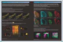
Channel unmixing Fast multiphoton imaging, powerful enough for in vivo imaging Channel unmixing The Nikon resonant scanner is capable of high-speed 420-fps imaging, the world’s fastest for a multiphoton microscope using point scanning technology. Unique to this design is a resonant scan mirror capable of imaging full fields of view at much higher speeds than traditional galvano scanners. Nikon's optical pixel clock system, which monitors the position of the resonant mirror in real time, adjusts the pixel clock to ensure more stable, geometrically correct and more evenly illuminated imaging even...
Open the catalog to page 4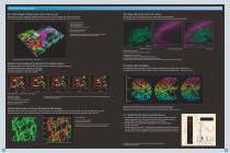
Channel unmixing Multiphoton imaging gallery Four-color imaging of human colon cancer cells in in vivo SHG image of the brain surface of a mouse Three-dimensional volume rendering of implanted subcutaneous tumor of HCT116 expressing Fucci. The cell cycle of tumor cells and the environment (collagen fiber and vessels) are visualized. Upper right, only collagen fiber and vessels are shown. The neocortex of an H-line 5-week-old mouse was studied with the open skull method. The SHG signals from dura mater and EYFP fluorescence signals were simultaneously acquired using the NDD. Red: Fucci mkO2/cancer...
Open the catalog to page 5
A1 MP+/A1R MP+ achieve the most advanced multiphoton imaging Standard NDD Nikon’s high-NA objectives are ideal for multiphoton imaging The fluorescence emissions from deep within a specimen are highly scattered in multiphoton excitation, and therefore the conventional detector using a pinhole cannot provide bright fluorescent images. The episcopic NDD in the A1 MP+/A1R MP+ is located close to the back aperture of the objective to detect the maximum amount of scattered emission signals from deep within living specimens. The use of this four-channel detector in combination with special spectral...
Open the catalog to page 6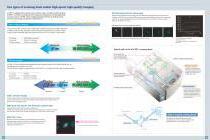
Two types of scanning head enable high-speed, high-quality imaging A1 MP+ is equipped with a galvano (non-resonant) scanner for high-resolution imaging. A1R MP+ is a hybrid scanning head that incorporates both galvano and ultrahigh-speed resonant scanners. A1R MP+ allows imaging and photoactivation at ultrafast speeds necessary for revealing cell dynamics and interaction. Ultrafast photoactivation and imaging High-resolution imaging Photoactivation during fluorescence imaging can be conducted using both galvano and resonant scanners. Two types of A1R MP+, the conventional 1080 nm type and the...
Open the catalog to page 7
Key Nikon innovations for improving image quality Enhanced spectral detector The best image quality is achieved by an increased light sensitivity resulting from comprehensive technological innovations in electronics, optics and software. Nikon’s original spectral performance is even further enhanced in the A1 MP+ series, allowing high-speed spectral acquisition with a single scan. In addition, advanced functions, including real-time unmixing, are incorporated. Low-angle incidence dichroic mirror With the A1 MP+ series, the industry’s first low-angle incidence method is utilized on the dichroic...
Open the catalog to page 8
Intuitive, easy-to-use software for multiphoton imaging NIS-Elements C Acquisition and Analysis software Simple operations common with Nikon confocal microscopes Channel unmixing function Microscope control •All necessary operations for image capture are displayed in one window. •Lasers and detectors for visible laser excitation can be switched simply by selecting fluorescent probe to be used. •One-touch switching of high speed resonant scanner and high-resolution galvano (non-resonant) scanner •Simultaneous photoactivation with high speed imaging is possible with visible laser excitation. Nikon's...
Open the catalog to page 9
System diagram ^ Laser Unit for Multiphoton Microscopy ^ Chameleon Vision II MaiTaiHP/eHP InSight DeepSee*1 Laser Unit for Confocal Microscopy ^ LU-LR 4-laser Power Source Rack ^ Detector Unit for Confocal Microscopy^ A1-DUS Spectral Detector Unit Filter Cubes ^ Incident Optical Unit ^ ^ Scan Head and Controller ^ Scan Head Detector Unit for Multiphoton Microscopy Diascopic Non-descanned Episcopic Non-descanned Detector (Standard/ (Standard/ Filter Cubes *1 When using a scan head for 1300 nm *2 When using Spectral Detector Unit. *3 Dedicated adapter is required depending on microscope model....
Open the catalog to page 10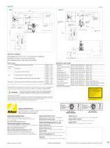
Laser Chiller for Multiphoton Microscopy Incident Optical Unit Incident Optical Unit Laser for Multiphoton Microscopy Laser Controller for Multiphoton Microscopy Laser Controller for Multiphoton Microscopy Scan Head Scan Head Non-Descanned Detector Vibration Isolated Table Non-Descanned Detector 4 Detector Unit, Spectral Detector Unit, Controller Vibration Isolated Table 4 Detector Unit, Spectral Detector Unit, Controller Laser Chiller for Multiphoton Microscopy 4-laser Module A, 4-laser Power Source Rack Approx. 2750 4-laser Module A, 4-laser Power Source Rack Laser for Multiphoton Microscopy...
Open the catalog to page 11All Nikon Instruments catalogs and technical brochures
 Super Resolution Microscopes
28 Pages
 HCA
HCA8 Pages
 DS-Fi3
16 Pages
 A1 HD25 / A1R HD25
15 Pages
 TI-E
TI-E32 Pages
 ECLIPSE TS2
8 Pages
 ECLIPSE TS2R
8 Pages
 N-SIME
6 Pages
 N-SIM N-STORM
24 Pages
 ECLIPSE E100 LED
8 Pages
 ECLIPSE Ti2
24 Pages
 A1 MP+ / A1R MP+
20 Pages
 DIGITAL SIGHT SERIES
16 Pages
 Eclipse LV100N-POL Ci-POL
8 Pages
 Eclipse Ti
32 Pages
 C2 Plus
16 Pages
 Biological Microscopes
24 Pages
 Stereomicroscopes
32 Pages
 SMZ18
SMZ1816 Pages
 N-SIM E
4 Pages


Archived catalogs
 A1 MP+ / A1R MP
15 Pages
 ShuttlePix
8 Pages
 Eclipse FN1
12 Pages
 NeoScope Brochure
16 Pages
 ShuttlePix Brochure
5 Pages
 Bio Station IMQ
8 Pages
 MiBrochure
2 Pages
 AZ_C1
AZ_C12 Pages
 High Content Microscopy
2 Pages
 C2+_2CE-SCHH-5
16 Pages
 A1+_A1R+_2CE-SBTH-9
13 Pages
 N-STORM 4.1
13 Pages
 A1+ / A1R+
13 Pages
 NIS-Elements
8 Pages
 Digital Sight Series
9 Pages
 AZ100M
8 Pages
 ShuttlePix P-400R
5 Pages
 Eclipse Ni
15 Pages
 Eclipse Ci-E/ Ci-L
5 Pages
 AZ100
AZ1004 Pages
 Eclipse E200POL
3 Pages
 Eclipse LV100ND POL/DS
2 Pages
 Eclipse E100 LED
4 Pages
 Eclipse E200 POL
3 Pages
 Biological Microscopes
12 Pages
 ECLIPSE LV100N POL 50i/ POL
4 Pages
 Eclipse E100-LED
8 Pages
 A1 MP+ / A1R MP+
11 Pages
 Digital Sight Series Brochure
16 Pages
- Compound microscope
- Laboratory microscope
- Desktop microscope
- Visualization software
- Tablet computer software
- Tablet PC software
- Control software
- Laboratory software
- CMOS camera
- Camera with USB port
- LED microscope
- LED light source
- Capture software
- Training software
- Biology microscope
- HD camera
- Microscope camera
- Research microscope