- Products
- Catalogs
- News & Trends
- Exhibitions
AZ_C1

AZ_C1
- Optical Zoom: AZ100 optical zoom ranges from 1-8x, while the C1 confocal scan zoom ranges from 1x-1000x.
- Focus: The system includes an EPI stand with an 85 mm focus range and a DIA stand with coarse and fine focus adjustments.
- Objectives: Various objective lenses are available, including AZ-Plan Apo and AZ-Plan Fluor, with different numerical apertures and working distances.
- Laser Light Source: Multiple laser wavelength options are available, including laser diodes, Ar lasers, and HeNe lasers, with varying power outputs.
- Fluorescence Detector: The system supports 2 to 3 channels, with display modes ranging from 160 x 160 to 2048 x 2048 pixels.
- Spectral Detector: Features 32 channels with a wavelength resolution of 2.5/5/10 nm and a minimum wavelength step of 0.2 nm.
- Power Requirements: The confocal system requires approximately 830 W.
- Macro to Micro Imaging: The AZ-C1 allows continuous imaging from macro to micro levels with a single microscope, using a combination of low and high magnification objective lenses and a scanning zoom function.
- High NA Objectives: These enable fast, high-resolution, single-image capture of wide specimen areas, suitable for observing embryos and cell populations.
- Time-Lapse Observation: The system supports time-lapse observation of dynamic cell behaviors.
- Deep Imaging: Capable of imaging deep into specimens, capturing fluorescence signals efficiently in both macro and micro imaging.
- C1plus: Standard model with high resolution, sensitivity, and contrast, suitable for single laboratories or large research groups.
- C1si-Ready: Upgradable to C1si by adding a spectral detector.
- C1si: Features a 32-channel multianode spectral detector, capturing a spectral bandwidth of 320 nm in a single scan.
Catalog excerpts
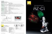
Macro confocal microscope system AZ-C1 Specifications AZ100 optical zoom C1 confocal scan zoom AZ-STE EPI stand/ AZ-STD DIA stand AZ-Plan Apo 0.5x (NA 0.05/W.D. 54 mm), AZ-Plan Apo 1x (NA 0.1/W.D. 35 mm), AZ-Plan Fluor 2x (NA 0.2/W.D. 45 mm), AZ-Plan Apo 4x (NA 0.4/W.D. 20 mm), AZ-Plan Fluor 5x (NA 0.5/W.D. 15 mm) Laser light source Laser wavelength options Focus range: 85 mm stroke Coarse focus: 18.5 mm/rotation Fine focus: 3.27 mm/rotation Stage focus: 10 mm range with 0.27 mm/rotation under software* control with remote focus accessory Laser diode (445 nm, 17 or 40 mW), Ar laser (488 nm, 10 mW), Ar laser (457, 477, 488 or 514 nm, 50 mW), Solid state laser (488 nm, 20 or 50 mW), G-HeNe laser (543 nm, 1.5 mW polarized), Solid state laser (561 nm, 10, 20 or 50 mW), Y-HeNe laser (594 nm, 2 mW), R-HeNe laser (633 nm, 5 mW), Laser diode (640 nm, 20 or 40 mW) Maximum number Laser control options Macro confocal microscope system Laser shutter Motorized mechanical shutter (each laser) fluorescence detector Display mode Scanning speed Standard: 1 fps (512 x 512 pixels), Bi-directional scanning: 1.4 fps (512 x 512 pixels) Corresponding wavelength Wavelength resolution Minimum wavelength step Display mode Power Scanning speed Standard: 0.5 fps (512 x 512 pixels, 32 channel simultaneous recording) Confocal system (PC, monitor, C1 controller, AOM controller): Approx. 830 W (single phase AC 115 V, 7.2 A / AC 230 V, 3.6 A, with earth) (Does not include microscopes and lasers.) Dimensional diagram (Unit: mm) TO ENSURE CORRECT USAGE, READ THE CORRESPONDING MANUALS CAREFULLY BEFORE USING YOUR EQUIPMENT. Product images appearing in this brochure do not include the laser safety stage cover. The diascopic detector is under development. Sample images in this brochure captured with the prototype only. AZ-C1 is a combination of the AZ100 microscope and the C1 series confocal laser microscope system. The AZ100M and A1 series are not compatible. Specifications and equipment are subject to change without any notice or obligation on the part of the manufacturer. September 2009 ©2009 NIKON CORPORATION NIKON CORPORATION 6-3, Nishiohi 1-chome, Shinagawa-ku, Tokyo 140-8601, Japan phone: +81-3-3773-8973 fax: +81-3-3773-8986 http://www.nikon.com/instruments/ NIKON INSTRUMENTS INC. 1300 Walt Whitman Road, Melville, N.Y. 11747-3064, U.S.A. phone: +1-631-547-8500; +1-800-52-NIKON (within the U.S.A. only) fax: +1-631-547-0306 http://www.nikoninstruments.com/ NIKON INSTRUMENTS EUROPE B.V. Laan van Kronenburg 2, 1183 AS Amstelveen, The Netherlands phone: +31-20-44-96-300 fax: +31-20-44-96-298 http://www.nikoninstruments.eu/ NIKON INSTRUMENTS (SHANGHAI) CO., LTD. CHINA phone: +86-21-5836-0050 fax: +86-21-5836-0030 (Beijing branch) phone: +86-10-5869-2255 fax: +86-10-5869-2277 (Guangzhou branch) phone: +86-20-3882-0552 fax: +86-20-3882-0580 NIKON SINGAPORE PTE LTD SINGAPORE phone: +65-6559-3618 fax: +65-6559-3668 NIKON UK LTD. UNITED KINGDOM phone: +44-208-247-1717 fax: +44-208-541-4584 NIKON MALAYSIA SDN. BHD. MALAYSIA phone: +60-3-7809-3688 fax: +60-3-7809-3633 NIKON GMBH AUSTRIA AUSTRIA phone: +43-1-972-6111-00 fax: +43-1-972-6111-40 NIKON INSTRUMENTS KOREA CO., LTD. KOREA phone: +82-2-2186-8410 fax: +82-2-555-4415 NIKON CANADA INC. CANADA phone: +1-905-602-9676 fax: +1-905-602-9953 NIKON FRANCE S.A.S. FRANCE phone: +33-1-4516-45-16 fax: +33-1-4516-45-55 NIKON BELUX BELGIUM phone: +32-2-705-56-65 fax: +32-2-726-66-45 NIKON GMBH GERMANY phone: +49-211-941-42-20 fax: +49-211-941-43-22 NIKON INSTRUMENTS S.p.A. ITALY phone: +39-055-300-96-01 fax: +39-055-30-09-93 NIKON AG SWITZERLAND phone: +41-43-277-28-67 fax: +41-43-277-28-61 Product images appearing in this brochure do not include the laser safety stage cover. The laser safety stage cover should be attached during actual use. This brochure is printed on recycled paper made from 40% used material.
Open the catalog to page 1
New system for high-definition macro confocal image acquisition Comparison of same specimen regions captured by the AZ-C1 and an epi-fluorescence microscope The AZ-C1 eliminates out-of-focus light and flare to deliver highly resolved confocal fluorescence images and optical sections. The AZ-C1 enables high-definition confocal image acquisition during macro observation. Sharp wide field of view images with unprecedentedly high S/N ratios allow for imaging of whole-mount specimens such as embryos and large tissue slices that are commonly used in developmental and systems biology studies. Moreover,...
Open the catalog to page 2All Nikon Instruments catalogs and technical brochures
 Super Resolution Microscopes
28 Pages
 HCA
HCA8 Pages
 DS-Fi3
16 Pages
 A1 HD25 / A1R HD25
15 Pages
 TI-E
TI-E32 Pages
 ECLIPSE TS2
8 Pages
 ECLIPSE TS2R
8 Pages
 N-SIME
6 Pages
 N-SIM N-STORM
24 Pages
 ECLIPSE E100 LED
8 Pages
 ECLIPSE Ti2
24 Pages
 A1 MP+ / A1R MP+
20 Pages
 DIGITAL SIGHT SERIES
16 Pages
 Eclipse LV100N-POL Ci-POL
8 Pages
 Eclipse Ti
32 Pages
 C2 Plus
16 Pages
 Biological Microscopes
24 Pages
 Stereomicroscopes
32 Pages
 SMZ18
SMZ1816 Pages
 N-SIM E
4 Pages


Archived catalogs
 A1 MP+ / A1R MP
15 Pages
 ShuttlePix
8 Pages
 Eclipse FN1
12 Pages
 NeoScope Brochure
16 Pages
 ShuttlePix Brochure
5 Pages
 Bio Station IMQ
8 Pages
 MiBrochure
2 Pages
 High Content Microscopy
2 Pages
 C2+_2CE-SCHH-5
16 Pages
 A1+_A1R+_2CE-SBTH-9
13 Pages
 N-STORM 4.1
13 Pages
 A1_MP
A1_MP11 Pages
 A1+ / A1R+
13 Pages
 NIS-Elements
8 Pages
 Digital Sight Series
9 Pages
 AZ100M
8 Pages
 ShuttlePix P-400R
5 Pages
 Eclipse Ni
15 Pages
 Eclipse Ci-E/ Ci-L
5 Pages
 AZ100
AZ1004 Pages
 Eclipse E200POL
3 Pages
 Eclipse LV100ND POL/DS
2 Pages
 Eclipse E100 LED
4 Pages
 Eclipse E200 POL
3 Pages
 Biological Microscopes
12 Pages
 ECLIPSE LV100N POL 50i/ POL
4 Pages
 Eclipse E100-LED
8 Pages
 A1 MP+ / A1R MP+
11 Pages
 Digital Sight Series Brochure
16 Pages
- Analysis software
- Compound microscope
- Laboratory microscope
- Desktop microscope
- Visualization software
- Tablet computer software
- Tablet PC software
- Control software
- LED microscope
- Laboratory software
- CMOS camera
- Camera with USB port
- LED light source
- Capture software
- Biology microscope
- Training software
- Microscope camera
- HD camera
- Binocular microscope