- Products
- Catalogs
- News & Trends
- Exhibitions
DS-Fi3
1 /16Pages

DS-Fi3
1 /16Pages
Catalog excerpts

Digital Cameras for Microscopes
Open the catalog to page 1
Nikon Digital Sight Series New Lineup A new system for imaging: the DS-Fi3, a high resolution and sensitivity general purpose color camera has been added to the Nikon Digital Sight series. The DS-Fi3 can be connected to a PC, or the new compact tablet-style DS-L4.
Open the catalog to page 2
Microscope Camera Highresolution Tubular adenoma, HE staining (Objective: CFI Plan Apochromat λ 4x) Photos courtesy of:Dr. Yasunori Ohta, Department of Pathology, IMSUT Hospital, Institute of Medical Science, The University of Tokyo High-resolution images A CMOS high density 5.9 megapixel sensor produces high resolution images. USB3.0 date transfer allows fast focusing at high resolution, and easy capture images in all types of observation methods such as brightfield, differential interference contrast, and phase contrast. Liquid crystal panel (Objective: TU Plan Fluor 10x) 3
Open the catalog to page 3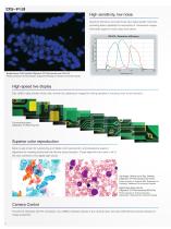
High sensitivity, low noise Quantum efficiency and read noise have been greatly improved, providing better capability for acquisition of fluorescent images with better signal-to-noise ratios than before. Quantum efficiency (%) Breast cancer, FISH method (Objective: CFI Plan Apochromat 100x Oil) Photos courtesy of: Hironao Kusakari, Diagnostic Pathology, St. Marianna University Hospital High-speed live display Fast USB3.0 data transfer means fast, smooth live updating of images for finding samples or focusing, even at full resolution. The mounting board (Objective: TU Plan Fluor 5x) Superior color...
Open the catalog to page 4
Microscope Camera Control Unit Compact, easy-to-use tablet-type microscope camera control unit. DS-Fi3 can be optionally connected to the DS-L4 tablet-style control unit, eliminating the need and space requirements of a desktop PC. DS-L4 has a large number of built-in functions for measurement and annotations, and has built-in security for network connectivity. Tablet-type camera control unit Large, 10.1 inch, touch-screen 1920 x 1200 pixel display: The DS-Fi3 can be set and operated simply and easily through the tablet by touch, or by connecting Bluetooth accessories such as a keyboard or mouse....
Open the catalog to page 5
Two Large Sensor high resolution 16.25-megapixel CMOS image sensors for microscopy Two Nikon FX-format CMOS image sensor cameras join the Digital Sight series of microscope digital cameras: the DS-Ri2 color digital camera and the DS-Qi2 monochrome digital camera. High pixel density and large field of view coupled with USB3.0 high speed data transfer offer fast frame rates and high resolution images with these CMOS image sensors. Large Format CMOS image sensors Nikon manufactures CMOS image sensors and imaging technologies for professional DSLR cameras, and now has optimized our sensors for microscopy...
Open the catalog to page 6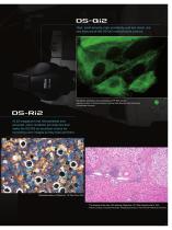
High pixel density, high sensitivity and low noise are key features of the DS-Qi2 monochrome camera. Pig kidney epithelial cells expressing GFP-EB3 tubulin Sample courtesy of: Michael Davidson, National High Magnetic Field Laboratory, Florida State University 16.25 megapixel (not interpolated) and accurate color rendition are features that make the DS-Ri2 an excellent choice for recreating color images as they eyes see them. Malleablecastiron (Objective: TU Plan Fluor 20x) The tissues of the liver, HE staining (Objective: CFI Plan Apochromat λ 10x) Photos courtesy of: Kazuhiro Muraoka, Photography...
Open the catalog to page 7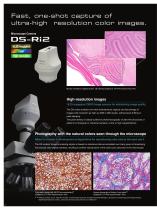
Fast, one-shot capture of ultra-high resolution color images. Microscope Camera 16.25 megapixel Color High-resolution Mouse cerebellum sagittal section, HE staining (Objective: CFI Plan Apochromat λ 4x) High-resolution images 16.25-megapixel CMOS image sensors for astonishing image quality The DS series enables one-shot instantaneous capture and fast storage of images with resolution as high as 4908 x 3264 pixels, without pixel shifting or pixel stepping. This pixel density is ideally suited for photomicrography of ultra-fine structures or patterns in biological or industrial samples, at low or...
Open the catalog to page 8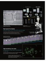
Conventional camera Semiconductors (IC wafers) (Objective: TU Plan Fluor 20x) Resolution chart (Objective: TU Plan Fluor 20x) High-speed live display High-speed display, even of supra-HDTV-class live images The DS-Ri2 can display 4908×3264 pixel (full-pixel) images at 6 fps, or 1636×1088 pixel (3×3 pixel averaging) images at 45 fps. This fastlive frame rate makes fine focusing easy to perform. Example of combination with the LV100ND industrial microscope Semiconductors (IC wafers) (Objective: TU Plan Fluor 5x) 1636×1088 pixel / Exposure time: 100μsec High sensitivity, low noise Fluorescent color...
Open the catalog to page 9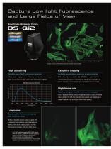
Capture Low light fluorescence and Large Fields of View Monochrome Microscope Camera 16.25 megapixel Monochrome Cooled Indian Muntjac Deer Skin Fibroblast Cells, Cytoskeletal F-actin labeled with Alexa Fluor 488 Sample courtesy of: Michael Davidson and Florida State University High sensitivity Excellent linearity 7.3μm pixels, high quantum efficiency, and very low read noise With a linearity error of ±1%, the DS-Qi2 is a superb tool for Detects even faint fluorescent signals Reliable quantitative analysis made possible allow the DS-Qi2 to read in even faint fluorescent signals. based intensity measurement...
Open the catalog to page 10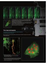
Time-lapse images (every 1 second) of LLC-PK1 cells with GFP-EB3 tubulin. Each image represents the maximum intensity projection of the timelapse, allowing visualization of the end-binding protein located on the microtubule plus-ends, and allowing tracing of the microtubule path. DS-Qi2 captures an extremely large field of view, but still represents very fine details as demonstrated in this cropped timelapse sequence from a large FOV image. Objective: CFI Plan Apochromat λ 60x oil / NA: 1.4) Sample courtesy of: Michael Davidson, National High Magnetic Field Laboratory, Florida State University...
Open the catalog to page 11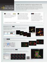
Imaging software Integration with the comprehensive imaging software series Nikon uses the NIS-Elements series as control software. NIS-Elements allows functions from basic imaging to control of the microscope and peripheral devices to be performed, as well as the measurement, analysis, and management of acquired images. Four basic packages and a variety of optional modules are available to suit every application and objective. * See the NIS-Elements Catalog for details. Free package The bundled free package offers functions for the display of scale on live images, full-screen display, and more....
Open the catalog to page 12All Nikon Instruments catalogs and technical brochures
 Super Resolution Microscopes
28 Pages
 HCA
HCA8 Pages
 A1 HD25 / A1R HD25
15 Pages
 TI-E
TI-E32 Pages
 ECLIPSE TS2
8 Pages
 ECLIPSE TS2R
8 Pages
 N-SIME
6 Pages
 N-SIM N-STORM
24 Pages
 ECLIPSE E100 LED
8 Pages
 ECLIPSE Ti2
24 Pages
 A1 MP+ / A1R MP+
20 Pages
 DIGITAL SIGHT SERIES
16 Pages
 Eclipse LV100N-POL Ci-POL
8 Pages
 Eclipse Ti
32 Pages
 C2 Plus
16 Pages
 Biological Microscopes
24 Pages
 Stereomicroscopes
32 Pages
 SMZ18
SMZ1816 Pages
 N-SIM E
4 Pages


Archived catalogs
 A1 MP+ / A1R MP
15 Pages
 ShuttlePix
8 Pages
 Eclipse FN1
12 Pages
 NeoScope Brochure
16 Pages
 ShuttlePix Brochure
5 Pages
 Bio Station IMQ
8 Pages
 MiBrochure
2 Pages
 AZ_C1
AZ_C12 Pages
 High Content Microscopy
2 Pages
 C2+_2CE-SCHH-5
16 Pages
 A1+_A1R+_2CE-SBTH-9
13 Pages
 N-STORM 4.1
13 Pages
 A1_MP
A1_MP11 Pages
 A1+ / A1R+
13 Pages
 NIS-Elements
8 Pages
 Digital Sight Series
9 Pages
 AZ100M
8 Pages
 ShuttlePix P-400R
5 Pages
 Eclipse Ni
15 Pages
 Eclipse Ci-E/ Ci-L
5 Pages
 AZ100
AZ1004 Pages
 Eclipse E200POL
3 Pages
 Eclipse LV100ND POL/DS
2 Pages
 Eclipse E100 LED
4 Pages
 Eclipse E200 POL
3 Pages
 Biological Microscopes
12 Pages
 ECLIPSE LV100N POL 50i/ POL
4 Pages
 Eclipse E100-LED
8 Pages
 A1 MP+ / A1R MP+
11 Pages
 Digital Sight Series Brochure
16 Pages