- Catalogs
- Nikon Instruments
- N-SIM N-STORM
- Products
- Catalogs
- News & Trends
- Exhibitions
N-SIM N-STORM
1 /24Pages

N-SIM N-STORM
1 /24Pages
Catalog excerpts
Super Resolution Microscope N-SIM/N-STORM Super Resolution Microscope
Open the catalog to page 1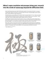
Nikon’s super-resolution microscopes bring your research into the world of nanoscopy beyond the diffraction limit. Nikon’s Super Resolution Microscope N-SIM/N-STORM enables elucidation of the structures and functions of nanoscopic machinery within living cells. The resolution of conventional optical microscopes, even with the highest numerical aperture optics, is limited by diffraction to approximately 200 nm. Using high-frequency structured illumination, the N-SIM can achieve an image resolution of 115 nm*, which was previously considered impossible with optical microscopes. Furthermore, with...
Open the catalog to page 2
Super Resolution Microscope See like you have never seen before
Open the catalog to page 3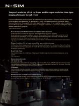
Temporal resolution of 0.6 sec/frame enables super-resolution time-lapse imaging of dynamic live cell events In structured illumination microscopy (SIM), the unknown cellular ultra-structure is elucidated by analyzing the moiré pattern produced when illuminating the specimen with a known high-frequency patterned illumination. Nikon’s Structured Illumination Microscope (N-SIM) realizes super resolution of up to 115 nm in multiple colors. In addition, it can continuously capture super-resolution images at a temporal resolution of 0.6 sec/frame*, enabling the study of dynamic interactions in living...
Open the catalog to page 4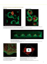
Double the resolution of conventional optical microscopes Volume view Maximum projection Width: 28.89 µm, Height: 27.83 µm, Depth: 17.20 µm Macrophages (J774 cells expressing mVenus-SNAP23) phagocytosing opsonized beads that were incubated with Alexa Fluor® 555 labeled secondary antibodies after fixation. The beads without red signals are in phagosomes containing mVenus-SNAP23. Photos courtesy of: Drs. Chie Sakurai, Kiyotaka Hatsuzawa and Ikuo Wada, Fukushima Medical University School of Medicine. Luminal surface of the organ of Corti at postnatal day 1. (Mouse) Green: F-actin, red: acetylated-tubulin...
Open the catalog to page 5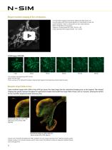
Super-resolution imaging of live cell dynamics Live-cell N-SIM imaging of mitochondria labeled with Mito-Tracker red. Live-cell imaging with N-SIM reveals dynamics of mitochondria at twice the spatial resolution. Cristae in mitochondria are also clearly observed. Mode: 3D-SIM (Slice reconstruction) Objective: CFI Apochromat TIRF 100XC Oil (NA 1.49) Image capturing interval: approximately 1 sec. (movie) N-SIM images (TIRF-SIM) FoLu cells (fox lung) expressing eGFP-vinculin Mode: TIRF-SIM mode Photos courtesy of: Dr. Michael W. Davidson, National High Magnetic Field Laboratory, Florida State University...
Open the catalog to page 6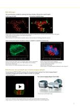
3D-SIM images Slice reconstruction is suitable for capturing time-lapse activities of living cells at specific depths. N-SIM image (3D-SIM) Conventional widefield image Bacillus subtilis bacterium stained with membrane dye Nile Red (red), and expressing the cell division protein DivIVA fused to GFP (green). N-SIM enables accurate localization of the protein during division. Reconstruction method: Slice Photos courtesy of: Drs Henrik Strahl and Leendert Hamoen, Centre for Bacterial Cell Biology, Newcastle University Stack reconstruction based on Gustafsson’s theory is suitable for acquisition...
Open the catalog to page 7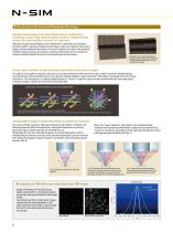
The principle of the Structured Illumination Microscopy Analytical processing of recorded moiré patterns, produced by overlaying a known high spatial frequency pattern, mathematically restores the sub-resolution structure of a specimen. Utilization of high spatial frequency laser interference to illuminate sub-resolution structures within a specimen produces moiré fringes, which are captured. These moiré fringes include modulated information of the sub-resolution structure of the specimen. Through image processing, the unknown specimen information can be recovered to achieve resolution beyond...
Open the catalog to page 8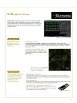
N-SIM analysis software N-SIM image processing, reconstruction and analysis are carried out using the N-SIM module that resides within Nikon’s universal, cross-platform imaging software NIS-Elements. The NIS-Elements platform allows for the same level of intuitive operation of N-SIM that exists for other Nikon imaging systems such as confocal microscopes. N-SIM image acquisition (3D-SIM) Image acquisition ・N-SIM mode selection ・Laser power control ・Setting imaging options ・JOBS option Setting image acquisition Up to five different laser wavelengths are available. User-customized spectral, z-stack,...
Open the catalog to page 9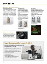
Objectives for super-resolution microscopes Silicone immersion objective Silicone immersion objective uses high viscosity silicone oil for immersion liquid, which has a refractive index that closely matches with those of the live cells. It allows capturing high-resolution, multi-color 3D images up to the apical side of a cell during long-term, time-lapse imaging. Superior chromatic aberration correction and high transmittance are ensured through broad wavelength range. Immersion objectives The adjustment and inspection of lenses using wavefront aberration measurement provide the SR objective...
Open the catalog to page 10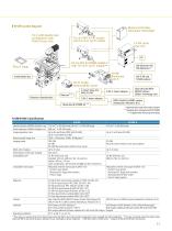
N-SIM system diagram Motorized HG fiber illuminator Intensilight Ti2-E with double layer configuration with Perfect Focus Unit TI2-LA-HTIRF H-TIRF module with TI2-LA-FL Epi-Fl module LU-NV series laser unit TI2-LA-NS2 N-STORM module 2 with TI2-LA-FL Epi-Fl module** Piezo Z stage N-SIM Shield box TI2-FT N-SIM motorizedfilter turret Vibration isolated table C-mount relay lens VM2.5X SIM Side port*** C-DA C-mount adapter Motorized N-STORM kit ** N-SIM illuminator unit R NIS-A 6D and N-SIM analysis iXon Ultra DU-897U EMCCD camera (Andor Technology Ltd.) ORCA-Flash4.0 sCMOS camera (Hamamatsu Photonics...
Open the catalog to page 11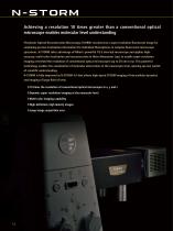
Achieving a resolution 10 times greater than a conventional optical microscope enables molecular level understanding STochastic Optical Reconstruction Microscopy (STORM) reconstructs a super-resolution fluorescent image by combining precise localization information for individual fluorophores in complex fluorescent microscope specimens. N-STORM takes advantage of Nikon’s powerful Ti2-E inverted microscope and applies highaccuracy, multi-color localization and reconstruction in three dimensions (xyz) to enable super-resolution imaging at tenfold the resolution of conventional optical microscopes...
Open the catalog to page 12All Nikon Instruments catalogs and technical brochures
 Super Resolution Microscopes
28 Pages
 HCA
HCA8 Pages
 DS-Fi3
16 Pages
 A1 HD25 / A1R HD25
15 Pages
 TI-E
TI-E32 Pages
 ECLIPSE TS2
8 Pages
 ECLIPSE TS2R
8 Pages
 N-SIME
6 Pages
 ECLIPSE E100 LED
8 Pages
 ECLIPSE Ti2
24 Pages
 A1 MP+ / A1R MP+
20 Pages
 DIGITAL SIGHT SERIES
16 Pages
 Eclipse LV100N-POL Ci-POL
8 Pages
 Eclipse Ti
32 Pages
 C2 Plus
16 Pages
 Biological Microscopes
24 Pages
 Stereomicroscopes
32 Pages
 SMZ18
SMZ1816 Pages
 N-SIM E
4 Pages


Archived catalogs
 A1 MP+ / A1R MP
15 Pages
 ShuttlePix
8 Pages
 Eclipse FN1
12 Pages
 NeoScope Brochure
16 Pages
 ShuttlePix Brochure
5 Pages
 Bio Station IMQ
8 Pages
 MiBrochure
2 Pages
 AZ_C1
AZ_C12 Pages
 High Content Microscopy
2 Pages
 C2+_2CE-SCHH-5
16 Pages
 A1+_A1R+_2CE-SBTH-9
13 Pages
 N-STORM 4.1
13 Pages
 A1_MP
A1_MP11 Pages
 A1+ / A1R+
13 Pages
 NIS-Elements
8 Pages
 Digital Sight Series
9 Pages
 AZ100M
8 Pages
 ShuttlePix P-400R
5 Pages
 Eclipse Ni
15 Pages
 Eclipse Ci-E/ Ci-L
5 Pages
 AZ100
AZ1004 Pages
 Eclipse E200POL
3 Pages
 Eclipse LV100ND POL/DS
2 Pages
 Eclipse E100 LED
4 Pages
 Eclipse E200 POL
3 Pages
 Biological Microscopes
12 Pages
 ECLIPSE LV100N POL 50i/ POL
4 Pages
 Eclipse E100-LED
8 Pages
 A1 MP+ / A1R MP+
11 Pages
 Digital Sight Series Brochure
16 Pages