
- Catalogs
- Nikon Instruments
- NeoScope Brochure

- Products
- Catalogs
- News & Trends
- Exhibitions
NeoScope Brochure

NeoScope Brochure
- Condenser Lens: Two-stage electromagnetic zoom condenser lens.
- Objective Lens: Electromagnetic lens.
- Auto Magnification: Corrected with reference to sample correction height.
- Preset Magnification: 6 levels, user programmable.
- Specimen Stage: Manual control for X and Y axes.
- Maximum Sample Size: 70 mm diameter × 50 mm height.
- Specimen Exchange: Draw-out mechanism.
- Image Memory: 1,280 × 960 × 16 bits.
- Pixels: 640 × 480, 1,280 × 960.
- Image Processing: Pixel and image accumulation.
- Automated Functions: Filament, alignment, focus, stigmator, exposure.
- Metrology: Distance between 2 points, angles.
- File Format: BMP, TIFF, JPEG.
- Computer: Desktop PC, OS Windows®7.
- Monitor: 23-inch wide LCD monitor (touch panel).
- Evacuation System: Fully automatic.
- Tilt Rotation Motorized Holder: Tilt: -15° to +45°; rotation: 360°.
- EDS: Available as an option.
- Voltage: Single phase AC 100 V (120 V, 220 V, 240 V).
- Frequency: 50/60 Hz.
- Power Consumption: Varies with voltage.
- Fluctuation: ±10% or less, with grounding.
- Room Temperature: 15 to 30°C.
- Humidity: 60% or less.
- Operation Table: Sturdy table with a loading capacity of 100 kg or more.
- Weight: Main console: approximately 50 kg.
- Base Unit Dimensions: 325 mm × 490 mm × 430 mm.
- NeoScope™
Catalog excerpts
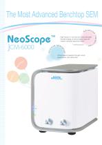
The Most Advanced Benchtop SEM High Vacuum or Low Vacuum mode selectable p> Sample imaging at various angles using a tilt "° rotation motorized specimen holder* ^ Utmost ease of operation through a touch panel system with intuitive GUI
Open the catalog to page 2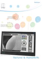
Speedy system startup A single touch to select High Vacuum or Low Vacuum mode Cartridge filament integrating wehnelt for easy exchange Auto gun alignment capability
Open the catalog to page 3
Sample: Iron rust Touch panel JEOL's renowned image quality can be achieved with the utmost ease. A touch panel system with intuitive GUI will guide the operator through the imaging process with the level of ease equal to a smartphone.
Open the catalog to page 4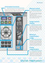
Automated functions Auto controls include alignment, focus, stigmator, contrast/ brightness, and Full Auto. A single touch of Full Auto will initiate the entire imaging process to present an image instantly. Manual control An item touched and selected will turn green. Hard, continued pressing of the buttons for coarse control. Light, intermittent tapping for fine control. These control buttons combines the ease of operation from the touch panel and the familiar feel of the knobs. Image search/ display The Display image button is used to search a library of images for a specific image. Checking...
Open the catalog to page 5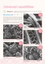
H S EV and Low Vacuum, allowing the mode Vacuum Observe A single touch can aswitch the images.between High I H operator to acquire variety of S EV I H S EV I HV BEI Secondary electron image in the High Vacuum mode can bring H out clear images of samples that require high magnification B EV image and samples with an uneven surface. Comparing SEI I H images with BEI images allows for closer examination of the B EV HV surface structure. I LV S may The image mode E also be switched to backscattered I compositional differences. H electron image to examine S EV LV I HV SEI LV HV HV BEI SEI 01 H V H...
Open the catalog to page 6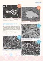
Sample: Aluminum alloy 10 μm Backscattered electron image Accelerating voltage 10 kV, magnification 3,000× Sample: Concrete Backscattered electron image Accelerating voltage 15 kV, magnification 200× H S EV I The Low Vacuum mode, a standard feature in NeoScope™, is designed to increase the pressure in the specimen chamber to neutralize the charges on the sample surface, allowing the operator to observe uncoated non conductive samples. The Low Vacuum mode is also effective for samples containing a small amount of oil or water and gas-emitting samples that resist coating. Sample: Filter paper...
Open the catalog to page 7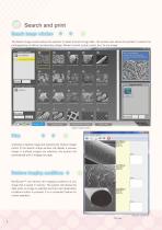
Qgffniil) ffimg& vjfiMto • The Search image window allows the operator to select and print image data. The window also allows the operator to restore the photographing conditions (accelerating voltage, filament current, probe current, etc.) for any image. Search image conditions Search image window Checking a desired image and pressing the Output images button in the Search image window will display a preview image. If multiple images are selected, the system will automatically print 3 images per page. NeoScope™ can retrieve the imaging conditions of any image that is saved in memory. The system...
Open the catalog to page 8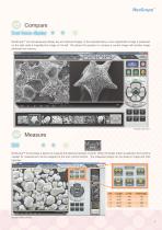
Compare NeoScope™ can simultaneously display live and retrieved images. In the example below, a low magnification image is presented on the right while a magnified live image on the left. This allows the operator to compare a current image with another image retrieved from memory. Sample: Star sand NeoScope™ incorporates a feature to measure the distance between 2 points. When the Scaler button is selected, the functions needed for measurement will be assigned to the auto control buttons. The measured results can be saved in image and CSV data files. Sample: Metal particles 9
Open the catalog to page 9
Elemental analysis Pressing the Analysis button will open the EDS view. EDS supports qualitative/quantitative analysis, point analysis, and mapping (elemental distribution). Sample: Black ore (mineral) Two buttons, Image and Spc, initiate analysis. Pressing the Quantitative button after data acquisition will display quantitative results. Pressing the Map button will initiate elemental mapping of the image. The mapped results will show 2 dimensional distribution of the constituent elements. Using mapped results, the operator can. Create quantitative maps in color where each color represents the...
Open the catalog to page 10
Analytical Assistance is one of the functions to help the operator navigate any data acquisition process, including mapping and line analysis. When a type of analysis is selected from the Analytical Assistance view, the system will display a series of process steps needed for the analysis. The buttons shown in the view will guide the operator through any analytical procedure. An example of mapping sequence When a line is defined on the image, the system will begin measuring relative concentration changes in the The elements may be edited after the stable analytical point
Open the catalog to page 11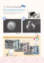
01 View at varying angles The Tilting and rotating motor drive holder allows the operator to examine samples at different angles. Observation of a tilted sample results in 3 dimensional information of the sample. The tilt rotation motorized specimen holder is optional. Sample: Substrate; magnification 45× Sample: Substrate; magnification 45× You can select any wallpaper for your GUI. Select one that you desire. JCM-6000 character Mr.Power Supply Sample: Spiderwort, uncoated, Low Vac mode
Open the catalog to page 12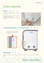
NeoScope™ will be ready for operation in 3 minutes after it is powered on. Placing a sample and closing the door will automatically initiate the evacuation sequence. An SEM image will be displayed when the evacuation is completed. A single touch on the panel can switch the mode between High Vacuum and Low Vacuum. NeoScope™ incorporates a JEOL proprietary dry SD detector. The detector is always ready for analysis during SEM imaging.
Open the catalog to page 13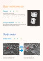
Changing filaments is simple and easy. Unlike the conventional filament assembly that requires cleaning of the wehnelt, the electron gun in NeoScope™ uses a pre-centered cartridge filament that integrates a wehnelt. Since the cartridge is replaced as a unit, cleaning of the wehnelt or centering of the filament is not needed. The exchange process is quick and insures correct positioning of the filament. Any new filament that is installed requires alignment to insure good image quality. This alignment process is fully automated in NeoScope™. Filament-wehnelt integral grid Coating allows non conductive...
Open the catalog to page 14All Nikon Instruments catalogs and technical brochures
 Super Resolution Microscopes
28 Pages
 HCA
HCA8 Pages
 DS-Fi3
16 Pages
 A1 HD25 / A1R HD25
15 Pages
 TI-E
TI-E32 Pages
 ECLIPSE TS2
8 Pages
 ECLIPSE TS2R
8 Pages
 N-SIME
6 Pages
 N-SIM N-STORM
24 Pages
 ECLIPSE E100 LED
8 Pages
 ECLIPSE Ti2
24 Pages
 A1 MP+ / A1R MP+
20 Pages
 DIGITAL SIGHT SERIES
16 Pages
 Eclipse LV100N-POL Ci-POL
8 Pages
 Eclipse Ti
32 Pages
 C2 Plus
16 Pages
 Biological Microscopes
24 Pages
 Stereomicroscopes
32 Pages
 SMZ18
SMZ1816 Pages
 N-SIM E
4 Pages


Archived catalogs
 A1 MP+ / A1R MP
15 Pages
 ShuttlePix
8 Pages
 Eclipse FN1
12 Pages
 ShuttlePix Brochure
5 Pages
 Bio Station IMQ
8 Pages
 MiBrochure
2 Pages
 AZ_C1
AZ_C12 Pages
 High Content Microscopy
2 Pages
 C2+_2CE-SCHH-5
16 Pages
 A1+_A1R+_2CE-SBTH-9
13 Pages
 N-STORM 4.1
13 Pages
 A1_MP
A1_MP11 Pages
 A1+ / A1R+
13 Pages
 NIS-Elements
8 Pages
 Digital Sight Series
9 Pages
 AZ100M
8 Pages
 ShuttlePix P-400R
5 Pages
 Eclipse Ni
15 Pages
 Eclipse Ci-E/ Ci-L
5 Pages
 AZ100
AZ1004 Pages
 Eclipse E200POL
3 Pages
 Eclipse LV100ND POL/DS
2 Pages
 Eclipse E100 LED
4 Pages
 Eclipse E200 POL
3 Pages
 Biological Microscopes
12 Pages
 ECLIPSE LV100N POL 50i/ POL
4 Pages
 Eclipse E100-LED
8 Pages
 A1 MP+ / A1R MP+
11 Pages
 Digital Sight Series Brochure
16 Pages
- Analysis software
- Compound microscope
- Laboratory microscope
- Desktop microscope
- Visualization software
- Tablet computer software
- Tablet PC software
- Control software
- Laboratory software
- CMOS camera
- Camera with USB port
- LED microscope
- LED light source
- Capture software
- Training software
- Biology microscope
- HD camera
- Microscope camera
- Research microscope