- Catalogs
- Nikon Instruments
- Super Resolution Microscopes
- Products
- Catalogs
- News & Trends
- Exhibitions
Super Resolution Microscopes
1 /28Pages

Super Resolution Microscopes
1 /28Pages
Catalog excerpts

Super Resolution Microscopes
Open the catalog to page 1
_N-SIM S_ Structured illumination microscopy 15 fps image acquisition Lateral resolution of -115 nm Axial resolution of ~269 nm
Open the catalog to page 2
The N-SIM S Super Resolution Microscope utilizes a unique high-speed structured illumination system to achieve acquisition speeds of up to 15 fps*, enabling fast biological processes to be captured at twice the spatial resolution of conventional light microscopes (~115nm** in XY). The N-STORM Super Resolution Microscope achieves a 10-fold improvement in resolution compared to conventional light microscopes (~20 nm in XY), enabling observation at the true molecular level. The N-SIM S and N-STORM can be easily combined within the same imaging system for greater flexibility in nanoscale imaging...
Open the catalog to page 3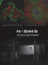
Lamellipodia of NG108 cell labeled with Alexa Fluor® 488 for actin (green) and TRITC-Phalloidin for microtubules (red). Photo courtesy of: Drs. Shizuha Ishiyama and Kaoru Katoh, The National Institute of Advanced Industrial Science and Technology (AIST) LLC-PK1 cell labeled with DAPI for nucleus (blue), Alexa Fluor® 488 for microtubules (green) and TRITC-Phalloidin for actin (red). Photo courtesy of: Dr. Kaoru Katoh, The National Institute of Advanced Industrial Science and Technology (AIST) See life in super resolution
Open the catalog to page 4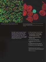
Synaptonemal complexes of C. elegans pachytene germ cells labeled with anti-SYP-1 antibodies. Photo courtesy of: Tyler Machovina and Dr. Judith Yanowitz, Magee-Womens Research Institute. Malaria parasite surface (MTIP) labeled with Alexa Fluor ® 488 (green), Erythrocyte membrane (Band 3) labeled with Alexa Fluor ® 568 (red), DNA labeled with DAPI (blue) Scientific Reports DOI:10.1038/s41598-018-22026-0 Photo courtesy of: Drs. Masayuki Morita, Eizo Takashima, Tadahiro Iimura, Takafumi Tsuboi, Proteo-Science Center, Ehime University The N-SIM S combines innovative structured illumination microscopy...
Open the catalog to page 5
Endosomes of COS7 cell labeled with YFP. Rapid movement of endosomes is captured at high resolution. Image acquisition speed: 6 fps Imaging mode: 3D-SIM Image courtesy of: Yasushi Okada, M.D., Ph.D., Department of Physics, Graduate School of Science, The University of Tokyo Scan the QR code to view a video illustrating super-resolution and widefield images. Capture rapid changes in live cells High-speed super-resolution imaging at 15 fps Nikon’s new high-speed structured illumination system utilizes a novel pattern modulation technology to generate fast and precise switching of illumination patterns....
Open the catalog to page 6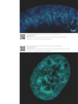
Growth cone of NG108 cell labeled with GFP-Lifeact for F-actin. Formation of actin mesh is captured at high-speed. Image acquisition speed: 10 fps Imaging mode: TIRF-SIM Image courtesy of: Drs. Minami Tanaka and Kaoru Katoh, The National Institute of Advanced Industrial Science and Technology (AIST) Scan the QR code to view a video illustrating super-resolution and widefield images. Histone H2B-GFP expressing in a HeLa cell. Visualization of fine movements of chromatin domains in different locations. Image acquisition speed: 3.9 fps Imaging mode: 3D-SIM Image courtesy of: Yuko Sato, Ph.D. and...
Open the catalog to page 7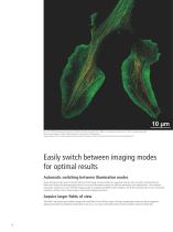
Two-color TIRF-SIM imaging of growth cone of NG108 cell labeled with Alexa Fluor ® 488 for F-actin (green) and Alexa Fluor ® 555 for microtubules (orange) Reconstructed image size: 2048 x 2048 pixels (66 μm x 66 μm with a 100X objective) Sample courtesy of: Drs. Shizuha Ishiyama and Kaoru Katoh, The National Institute of Advanced Industrial Science and Technology (AIST) Easily switch between imaging modes for optimal results Automatic switching between illumination modes Newly-developed, high-speed structured illumination technology not only enables fast acquisition rates but also automatic switching...
Open the catalog to page 8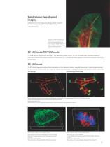
Simultaneous two-channel imaging Simultaneous two-color imaging is possible by utilizing an optional Two Camera Imaging Adaptor* and two sCMOS cameras. * Andor Technology Ltd. Growth cone of NG108 cell expressing GFP-LifeAct (F-actin, green) and mCherry-tubulin (microtubules, red) Photo courtesy of: Dr. Kaoru Katoh, The National Institute of Advanced Industrial Science and Technology (AIST) 2D-SIM mode/TIRF-SIM mode This mode captures super-resolution 2D images at high speed with incredible contrast. The TIRF-SIM mode enables Total Internal Reflection Fluorescence observation at double the resolution...
Open the catalog to page 9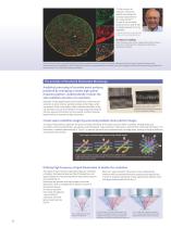
“N-SIM provides the resolution necessary to identify and evaluate the structural organization of the nuclear lamina*1, *2. Its ease of use and stable performance has made N-SIM an integral research tool in my laboratory.” *1 ol Biol Cell. 2015 Nov 5; 26(22):4075-86. M *2 Nature . 2017 Mar 9; 543(7644):261-264. Dr. Robert D. Goldman Ellison Foundation Senior Scholar, Stephen Walter Ranson Professor, Chair, Dept. of Cell & Mol. Biol., Feinberg School of Medicine, Northwestern University Lamin B1 (red) and Lamin C (green) form separate but interacting meshworks within the lamina of the embryonic...
Open the catalog to page 10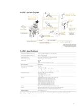
Ti2-E with double layer configuration with Perfect Focus Unit Motorized HG fiber illuminator Intensilight TI2-LA-HTIRF H-TIRF module with TI2-LA-FL Epi-Fl module TI2-LA-NS2 N-STORM module 2 with TI2-LA-FL Epi-Fl module** TI2-FT N-SIM motorizedfilter turret LU-NV series laser unit ORCA-Flash4.0 sCMOS camera (Hamamatsu Photonics K.K.) * Required when used with confocal system * Required when configured with N-STORM *** Supplied with microscope main body N-SIM S Specifications Lateral resolution (FWHM of beads in xy) *1 These values are measured using 100 nm diameter beads excited by a 488 nm laser....
Open the catalog to page 11
a: Hippocampal neurons and glia in culture b: Growth cone of a neuron in culture Experience the nanoscale universe
Open the catalog to page 12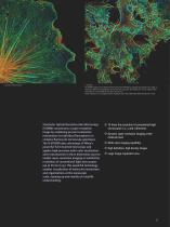
c: Glia in a neuronal culture d: COS cells 3D-STORM imaging of actin labeled with Alexa Fluor ® 647 Phalloidin using depth-code pseudo color. Image “a” shows four types of actin organization, from bottom left to top right: the cell body of a neuron, a glial cell with stress fibers, a neuronal dendrite with spines, and an axon. Photos courtesy of: Dr. Christophe Leterrier, NeuroCyto team, NICN CNRS-AMU UMR7259, Marseille, France STochastic Optical Reconstruction Microscopy (STORM) reconstructs a super-resolution image by combining precise localization information for individual fluorophores in...
Open the catalog to page 13All Nikon Instruments catalogs and technical brochures
 HCA
HCA8 Pages
 DS-Fi3
16 Pages
 A1 HD25 / A1R HD25
15 Pages
 TI-E
TI-E32 Pages
 ECLIPSE TS2
8 Pages
 ECLIPSE TS2R
8 Pages
 N-SIME
6 Pages
 N-SIM N-STORM
24 Pages
 ECLIPSE E100 LED
8 Pages
 ECLIPSE Ti2
24 Pages
 A1 MP+ / A1R MP+
20 Pages
 DIGITAL SIGHT SERIES
16 Pages
 Eclipse LV100N-POL Ci-POL
8 Pages
 Eclipse Ti
32 Pages
 C2 Plus
16 Pages
 Biological Microscopes
24 Pages
 Stereomicroscopes
32 Pages
 SMZ18
SMZ1816 Pages
 N-SIM E
4 Pages


Archived catalogs
 A1 MP+ / A1R MP
15 Pages
 ShuttlePix
8 Pages
 Eclipse FN1
12 Pages
 NeoScope Brochure
16 Pages
 ShuttlePix Brochure
5 Pages
 Bio Station IMQ
8 Pages
 MiBrochure
2 Pages
 AZ_C1
AZ_C12 Pages
 High Content Microscopy
2 Pages
 C2+_2CE-SCHH-5
16 Pages
 A1+_A1R+_2CE-SBTH-9
13 Pages
 N-STORM 4.1
13 Pages
 A1_MP
A1_MP11 Pages
 A1+ / A1R+
13 Pages
 NIS-Elements
8 Pages
 Digital Sight Series
9 Pages
 AZ100M
8 Pages
 ShuttlePix P-400R
5 Pages
 Eclipse Ni
15 Pages
 Eclipse Ci-E/ Ci-L
5 Pages
 AZ100
AZ1004 Pages
 Eclipse E200POL
3 Pages
 Eclipse LV100ND POL/DS
2 Pages
 Eclipse E100 LED
4 Pages
 Eclipse E200 POL
3 Pages
 Biological Microscopes
12 Pages
 ECLIPSE LV100N POL 50i/ POL
4 Pages
 Eclipse E100-LED
8 Pages
 A1 MP+ / A1R MP+
11 Pages
 Digital Sight Series Brochure
16 Pages
- Analysis software
- Compound microscope
- Laboratory microscope
- Desktop microscope
- Visualization software
- Tablet computer software
- Tablet PC software
- Control software
- LED microscope
- Laboratory software
- CMOS camera
- Camera with USB port
- LED light source
- Capture software
- Biology microscope
- Training software
- Microscope camera
- HD camera
- Binocular microscope