
- Catalogs
- Ocular Instruments
- Ocular Mainster Wide Field Laser Lens

Ocular Mainster Wide Field Laser Lens

Ocular Mainster Wide Field Laser Lens
The lens is designed for use by licensed physicians to counteract the optical power of the cornea, allowing for internal eye examination. Methylcellulose or similar fluids are recommended for lubrication and optical coupling. Precision optics refine the optical path and laser power density for treating fundus abnormalities. The laser spot magnification factor is crucial for calculating changes in laser spot size, and the stated laser spot size limit should not be exceeded for safety.
Design Features
The Ocular Mainster Wide Field Laser Lens is used for panretinal photocoagulation, providing an inverted aerial image of the retina at 0.68x magnification. It ensures distortion-free laser beam transmission and offers a binocular image across the entire field of view. The Mainster Wide Field EX Laser Lens offers an improved fundus image over a 145° field of view.
Technique
Familiarity with the lens is required. Use the slit lamp with parallel illumination and observation arms, and a vertical slit beam to minimize back-scattered light. Slit lamp magnification should be between 5x and 12x. The lens should be tilted on the cornea to select the viewing area, and the slit lamp must be moved further back compared to conventional lenses.
Product Specifications
Product codes include OMRA-WF and OMRA-WFEX, with static and dynamic fields of view ranging from 118° to 157°. The lens is designed by Martin A. Mainster, Ph.D., M.D., and features a contact diameter of 15.5mm and lens height of 28mm.
Warnings
Do not use the lens if damaged. It must be cleaned and sterilized before use, especially if in contact with an ulcerated cornea. Laser spot settings greater than 400 microns are not recommended to avoid excessive energy to the crystalline lens.
Contraindications
The lens is intended for transient use only and should not remain on the eye for prolonged periods. It must be sterilized if it has contacted an ulcerated cornea.
Contact Information
Ocular Instruments, 2255 116th Ave NE, Bellevue, Washington 98004-3039 USA. Phone: 425-455-5200 or 800-888-6616. Email: [email protected]. Website: www.ocularinc.com. Revised on 2017-08-23.
Catalog excerpts
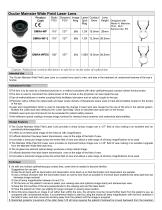
Ocular Mainster Wide Field Laser Lens Product Code Static Dynamic Image Laser Contact Lens FOV Mag Spot Diam Height Designed with: FOV Martin A. Mainster, Mag Ph.D., M.D. Kansas City, KS Caution: Federal law restricts this device to sale by or on the order of a physician. Intended Use The Ocular Mainster Wide Field Laser Lens is a contact lens used to view, and aids in the treatment of, anatomical features of the eye’s eye fundus. Indications for Use § The lens is to be used by a licensed physician in a method consistent with other ophthalmoscopic contact indirect fundus lenses. § The lens is used to counteract the optical power of the cornea so the physician can see inside the eye. § Use of methylcellulose or similar coupling fluids facilitates lubrication and an optical couple to the eye. § Precision optics refines the optical path and laser power de density of therapeutic lasers used to treat abnormalities located in the fundus of the eye. § Laser spot magnification factor is used to calculate the change in laser spot size caused by the use of the lens in the optic optical system. Multiply the Laser spot size setting etting by the “Laser Spot Mag” value to calculate laser spot size on the fundus. § Stated Laser spot size limit should not be exceeded for patient safety reasons. § Anti-reflective optical coatings increase ase image contrast for viewing ffundus anatomy and anatomical abnormalities. Design Features § The Ocular Mainster Wide Field Laser Lens provides a sharp fundus image over a 127°° field of view making it an excellent lens for panretinal photocoagulation. § It offers an inverted aerial image of the retina at .68x magnification. § It affords distortion free laser beam transmission, even at the edge of the field of view. § It provides a binocular image across the entire field of view and allows a wide range of sli slitt lamp magnifications to be used. § The Mainster Wide Field EX Laser Lens provides an improved fundus image over a 145 145°° field of view making it an excellent upgrade from the Mainster Wide field laser lens. § The all glass two element optical design produces a sharp retinal image. § It affords distortion free laser beam transmission, even at the edge of the field of view. § It provides a binocular image across the entire field of view and allows a wide range of slit lamp magnifications to be used. 4 Technique § As with any indirect ophthalmoscopy contact lens, some time is needed to become familiar. Suggestions for use are: § Use the slit lamp with its illumination and observation arms lined up so that illumination and observation are parallel. § Use a vertical slit beam with the illumination beam as narrow and short as possible to minimize back back-scattered scattered slit lamp light that can decrease image contrast. § Use slit lamp magnification between 5x and 12x. § Tilt the lens on the patient’s cornea to select your viewing area and optimize image clarity and stereoscopic view. § Keep the front surface of the lens perpendicular to the viewing axis and the laser beam. § Have the patient turn their eye slightly for larger changes in viewing area location. § Since this lens presents an image in air rather than within the lens, the slit lamp must be moved further back from the patie patient’s eye, as compared with conventional lenses. It is sometimes helpful to start by using the lowest slit lamp magnification with tthe lens centered in the field of view, and then move the slit lamp away from the patient until the image is acquired. § Restricted posterior movement of the older Zeiss 125 slit lamp requires the patient’s forehe
Open the catalog to page 1
This positioning may be achieved by placing a one inch thick sponge strip between the patient’s forehead and the headrest. Warning § Do not use if there are fractures, chips, scratches or other damage to the lens. § Lens must be properly cleaned and disinfected or sterilized before use. § If lens has been in contact with an ulcerated cornea it must be sterilized prior to subsequent use. Warning English: To avoid excessive energy to the crystalline lens, laser spot settings of greater than 400 microns are not recommended. Bulgarian: За да избегнете излишно подаване на енергия към лещата, не се...
Open the catalog to page 2All Ocular Instruments catalogs and technical brochures
 Product Catalog
82 Pages
 Ocular Imaging Eye Model
2 Pages
 OROCTS
1 Page
 Vitreolysis Sets
1 Page
- Anatomy model
- Training anatomy model
- Sterilization container
- Medical suitcase
- Sterilization tray
- Medical instruments support
- Eye anatomical model
- Custom medical suitcase
- Anatomical model kit
- Plastic medical suitcase
- Operating room support
- Medical imaging anatomical model
- Health education anatomical model
- Model holder
- Autoclavable sterilization tray
- Autoclavable sterilization container
- Lens sterilization tray