
- Catalogs
- Optopol Technology
- REVO 60

REVO 60
REVO 60
The document introduces the REVO 60, a state-of-the-art Spectral Domain Optical Coherence Tomography (OCT) device that simplifies eye examinations with advanced AI denoising technology and a user-friendly interface.
The REVO 60 utilizes AI algorithms to enhance tomogram quality, achieving results comparable to multiple averaged scans, thus improving early disease detection.
The REVO 60 combines multiple functionalities into one device, allowing comprehensive eye examinations from the retina to the cornea. It supports various operator and patient positions and requires minimal space for installation.
A single 3D retina scan performs both retina and glaucoma analysis, automatically recognizing eight retinal layers for precise diagnosis. The device offers high-density scanning and blood vessel recognition for accurate follow-up and progression tracking.
The REVO 60 supports DICOM and EMR integration, enhancing productivity and patient experience by allowing examination management across multiple stations and facilitating patient education.
The device offers non-invasive angiography for visualizing retinal microvasculature and widefield scans for comprehensive retina screening. It includes advanced imaging modes for detailed observation.
The REVO 60 provides tools for glaucoma diagnosis and monitoring, including nerve fiber layer and ganglion layer analysis. The Structure & Function report combines visual field and retinal data for comprehensive assessment.
The device offers detailed corneal curvature maps and keratoconus classification, utilizing various maps and display options for precise corneal condition assessment.
The REVO 60 allows anterior segment imaging without additional lenses and provides biometry parameters for ocular structure measurement, supporting IOL calculations with an up-to-date database.
The document provides detailed technical specifications, including light source, scanning speed, resolution, connectivity options, and power requirements.
Catalog excerpts
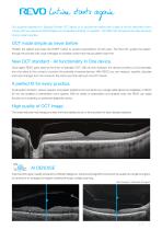
lution starts again Our supreme experience in Spectral Domain OCT allows us to provide the market with a state of the art instrument which comes with new advanced technologies and remarkable simplicity of operation. The REVO 60 will expand the daily demands of any modern practice. OCT made simple as never before Position the patient and press the START button to acquire examinations of both eyes. The Revo 60, guides the patient through the process with vocal messages to increase comfort and reduce patient chair time. New OCT standard - All functionality In One device. Once again REVO goes beyond...
Open the catalog to page 2
A single 3D Retina scan performs both Retina and Glaucoma analysis. The software automatically recognizes 8 retinal layers which assists with precise diagnosis and mapping of any changes in the patient’s condition. A variety of result analysis and presentation methods allows the most suitable selection to increase efficiency. FOLLOW-UP REVO’s standard high density scanning capability and blood vessel structure recognition enable a precise alignment of past and current scans. The operator can analyse changes in morphology, quantified progression maps and evaluate the progression trends. Progression...
Open the catalog to page 3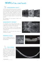
lution continues ANGIOGRAPHY SOCT1 This non-invasive dye free technique allows the visualization of the microvasculature of the retina. Both blood flow and structural visualization give additional diagnostic information about many retinal diseases. Angiography scan allows assessment of the structural vasculature of the macula, the periphery or the optic disc. Extremely short scanning times of 1,6 seconds in standard resolution or 3 seconds in high resolution. Now Angiography OCT can become a routine in your diagnostic practice. Mosaic mode: 10x6 mm ANGIOGRAPHY MOSAIC The Angiography mosaic delivers...
Open the catalog to page 4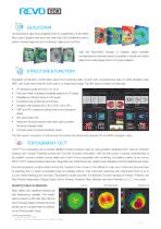
GLAUCOMA Comprehensive glaucoma analytical tools for quantification of the Nerve Fiber Layer, Ganglion layer and Optic Head with DDLS enable the user to perform precise diagnosis and monitoring of glaucoma over time. With the Asymmetry Analysis of Ganglion layers between hemispheres and between eyes it is possible to identify and detect glaucoma in early stages and in non-typical patients. STRUCTURE & FUNCTION2 Invaluable combination of information about the functional quality of vision with comprehensive data on retinal Ganglion Cells, RNFL and Optic Nerve Head for both eyes on a single report...
Open the catalog to page 5
lution continues ANTERIOR CHAMBER Built-in anterior lens allows the user to perform the imaging of the anterior segment without installing additional lens or forehead adapter. Now you can display the whole anterior segment or focus on a small area to bring out the details of the image. Anterior Chamber exam with a fast view of the whole Anterior Chamber make the evaluation of gonioscopy situation and the verification of cataract lens easier and faster. ANTERIOR For all anterior examination, no additional lens is required. This allows the examiner to quickly complete the scanning procedure. *...
Open the catalog to page 6
Choroidal observation Wide Central scan Sclera and Anterior Structure Cornea wide scan
Open the catalog to page 7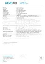
TECHNICAL SPECIFICATION Technology Light source Bandwidth Scanning speed Axial resolution Transverse resolution Overall scan depth Focus adjustment range Scan range Scan types Fundus image Alignment method Retina analysis Angiography OCT1 Angiography mosaic Glaucoma analysis Biometry OCT1 IOL Calculator3,4 Corneal Topography Map1 Anterior (no lens/adapter required) Connectivity Fixation target Dimensions (W×D×H) / Weight Power supply / consumption Spectral Domain OCT SLED, wavelength 850 nm 50 nm half bandwidth 60 000 measurements per second 2.6 μm digital, 5 μm in tissue 12 μm, typical 18 μm...
Open the catalog to page 8All Optopol Technology catalogs and technical brochures
 REVO NX 130
16 Pages
 REVO 80
8 Pages
 REVO FC 130
16 Pages
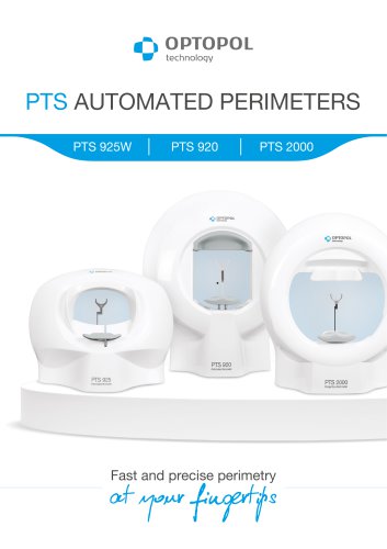 PTS AUTOMATED PERIMETERS
8 Pages
Archived catalogs
 revo-fc130
16 Pages
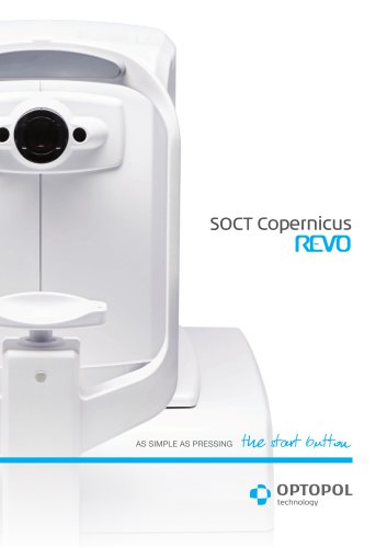 SOCT Coperncius REVO
6 Pages
 REVO FC
6 Pages
 REVO NX
12 Pages

- Analysis software
- Radiology software
- Hospital software
- Optopol fixed ophthalmic examination
- Automated software
- Traceability software
- Capture software
- Software module
- AI-assisted software
- Artificial intelligence software
- Import software
- 3D software
- Ophthalmoscope
- Screening software
- Analysis software module
- Medical software module
- Retinal camera
- Medical imaging software module
- Management software module
- Ophthalmic software