
- Catalogs
- Optopol Technology
- REVO FC 130

REVO FC 130
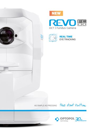
REVO FC 130
The document introduces two advanced ophthalmic devices: the REVO FC130 and the IOL Calculator and Biometry OCT2 systems. These devices integrate cutting-edge technologies to enhance diagnostic precision and patient comfort during eye examinations.
REVO FC130 Specifications and Features
The REVO FC130 is equipped with AccuTrackTM for real-time eye tracking and iTracking technology for patients with fixation difficulties. It combines OCT and fundus camera functionalities, providing comprehensive eye examinations from the cornea to the retina.
Ease of Use
The device features a single start button and voice-guided instructions, reducing patient chair time. Its compact design is suitable for small exam rooms, functioning as both a screening and diagnostic tool.
Imaging Capabilities
The REVO FC130 includes a 12.3 MP fundus camera with a 45° viewing angle and advanced image processing tools. It supports various view modes, including time comparisons of fundus photos.
Advanced Technologies
AI DenoiseTM improves tomogram quality by filtering noise, while Full Range technology provides extended depth scans. Motion Correction TechnologyTM ensures artifact-free images.
Networking and Integration
The device supports DICOM and EMR integration, enhancing connectivity with hospital systems and practice management software.
Glaucoma and Retina Analysis
Comprehensive glaucoma analysis tools include optic nerve parameters and ganglion layer analysis, with detailed reports for glaucoma management.
Angiography and Topography
The non-invasive Angiography A-OCT2 visualizes retinal microvasculature, and the Topography OCT2 module provides detailed corneal curvature maps.
IOL Calculator and Biometry OCT2 Systems
The IOL Calculator uses various formulas for IOL implant parameters and supports the latest IOL database standard. The Biometry OCT2 provides a complete set of biometry parameters and is available in Standard and Basic options.
Measurement Verification
The system allows visual verification of measurements with calipers, aiding in precise corrections with a 5 μm axial resolution.
OCT Gonioscopy and Anterior Chamber Scans
OCT gonioscopy visualizes iridocorneal angles and iris configuration, offering full-range anterior chamber scans for IOL position verification.
Fundus Camera and OCT Specifications
The non-mydriatic fundus camera captures color photographs with a 45° angle of view. The Spectral Domain OCT technology features a scanning speed of 130,000 measurements per second.
Retina and Glaucoma Analysis
Detailed retina analysis includes thickness measurements and angiography, with comprehensive glaucoma analysis.
Connectivity and Power
The system supports DICOM connectivity and operates within a power supply range of 100 V to 240 V.
Conclusion
The REVO FC130 and IOL Calculator and Biometry OCT2 systems are versatile, all-in-one devices that enhance diagnostic capabilities in ophthalmology, suitable for a wide range of clinical applications.
Catalog excerpts

REAL TIME EYE TRACKING
Open the catalog to page 1
OCT MADE SIMPLE AS NEVER BEFORE Once again REVO goes beyond the limits of standard OCT. With its new software, REVO enables full functionality from the cornea to the retina, combining the potential of several devices. With just a single OCT device you can measure, quantify, calculate and track changes from the cornea to the retina over time. A PERFECT FIT FOR EVERY PRACTICE Multiple Functions in One Device The REVO FC130 is an All in One device you can use in a number of ways such as, a full color Fundus Camera or as a combo providing simultaneous OCT and fundus images for high quality OCT imaging,...
Open the catalog to page 2
lution continues iTracking™ technology scans twice to compensate for any involuntary eye movements and blinks. It can be used for patients who have difficulty keeping their head on the chinrest during scanning with hardware eye tracking. After scanning the system immediately creates an artifact-free MC examination using the Motion Correction TechnologyTM. The elimination of eye movement and blinking artifacts ensures the high quality of Angio OCT images without patient inconvenience. Clear A-OCT data sets make it easier to interpret the condition of the retina vasculature. Improved tomogram quality...
Open the catalog to page 3
FUNDUS CAMERA A 12.3 MP Fundus Camera is integrated into our All in One OCT device capable of capturing detailed color images of ultra-high quality. The REVO FC130 is fully automated, safe and easy to use. Color fundus image capture is possible with a pupil as small as 3.3 mm. Easy to use fundus image processing tools deliver a stunning retinal image. Available modes deliver detailed photos of one or both eyes, as well as a chronological comparison of the fundus photos. Link a single fundus photo to several OCT scans. IR fundus preview and photo capture settings are adjusted automatically based...
Open the catalog to page 4
lution continues RETINA A single 3D Retina examination is sufficient to perform both Retina and Glaucoma analysis based on retinal scans. During the analysis, the software automatically recognizes eight retina layers to ensure a more precise diagnosis and mapping of any changes in the patient’s retina condition. 3D Single View Both View The high density of standard 3D scans allow the operator to precisely track disease progression. The operator can analyze changes in morphology, quantified progression maps and evaluate the progression trends. PRECISE REGISTRATION The software can track 3D scans...
Open the catalog to page 5
With the gold standard 14 optic nerve parameters and a new Rim to Disc and Rim Absence, the description of ONH condition is quick and precise. Advanced view provides combined information from Retina and Disc scans to integrate details of the Ganglion cells, RNFL, ONH in a wide field perspective for comprehensive analysis for both eyes. Ganglion Both Ganglion Progression The REVO DDLS (Disc Damage Likelihood Scale) uses 3 separate classifications for small, average and large discs. It supports the practicioner in a quick and precise evaluation of the patient’s glaucomatous disc damage. Asymmetry...
Open the catalog to page 6
lution continues COMPREHENSIVE GLAUCOMA SOLUTION1 Structure & Function - Combined OCT and VF results analysis Comprehensive glaucoma analytical tools for quantification of the Nerve Fiber Layer, Ganglion layer and Optic Nerve Head with DDLS provide precise diagnostics and monitoring of glaucoma over time. With the gold standard 14 optic nerve parameters and a new Rim to Disc and Rim Absence the description of ONH condition is quick and precise. COMPRHENSIVE STRUCTURE AND FUNCTION REPORT INCLUDES THE FOLLOWING: VF sensitivity results (24-2/30-2 or 10-2) • Total and Pattern Deviation probability...
Open the catalog to page 7
This non-invasive dye free technique allows the visualization of the microvasculature of the retina. Both blood flow and structural visualization give additional diagnostic information about many retinal diseases. Angiography scan allows assessment of the structural vasculature of the macula, the periphery or the optic disc. Extremely short scanning times of 1.6 seconds in standard resolution or 3 seconds in high resolution. Now, Angiography OCT can become routine in your diagnostic practice. ANGIO ANALYSIS METHODS Vessel Density Map The quantification tool provides quantification of the vasculature...
Open the catalog to page 8
lution continues A COMPLETE SET OF ANGIO OCT ANALYSIS VIEWS The software allows the user to observe, track and compare changes in the microvasculature of the retina in both eyes. Standard Single View Detailed Single View ANGIOGRAPHY MOSAIC The Angiography mosaic delivers high-detail images over a large field of the retina. Available modes present a predefined region of the retina in a convenient way. In manual mode it is possible to scan the desired region. Built-in analytics allow the user to see vascular layers, enface or thickness maps. Healthy patient, Angio Mosaic mode: 7×7 mm PDR, Angio...
Open the catalog to page 9
T-OCT™ is a pioneering way to provide detailed corneal curvature maps by using posterior dedicated OCT. Anterior, Posterior surfaces and Corneal Thickness provide the True Net Curvature information. With the Net power, a precise understanding of the patient’s corneal condition comes easily and is free of errors associated with modeling of posterior surface of the cornea. The REVO T-OCT module provides Axial maps, Tangential maps, Total Power map, Height maps, Epithelium and Corneal thickness maps. The corneal topography module shows the changes in the cornea on the difference map view. Customize...
Open the catalog to page 10
lution continues ANTERIOR CHAMBER The built-in anterior lens allows the user to perform imaging of the anterior segment without installing additional lenses or a forehead adapter. Now you can display the entire anterior segment or focus on a small area to bring out the details of the image. FULL RANGE TECHNIQUE Anterior Chamber exam with a fast view of the entire Anterior Chamber makes the evaluation of gonioscopy and the verification of cataract lens easier and faster. OCT gonioscopy provides the visualization of both iridocorneal angles together with information on iris confi guration on a...
Open the catalog to page 11All Optopol Technology catalogs and technical brochures
 REVO NX 130
16 Pages
 REVO 80
8 Pages
 REVO 60
8 Pages
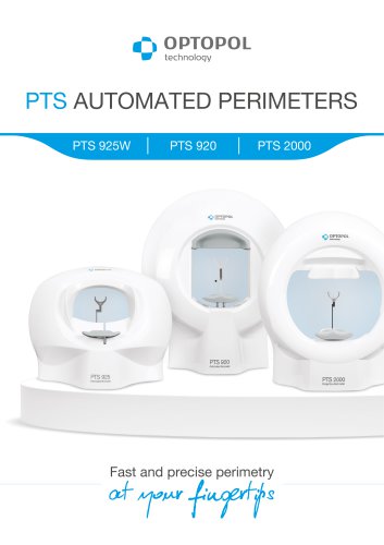 PTS AUTOMATED PERIMETERS
8 Pages
Archived catalogs
 revo-fc130
16 Pages
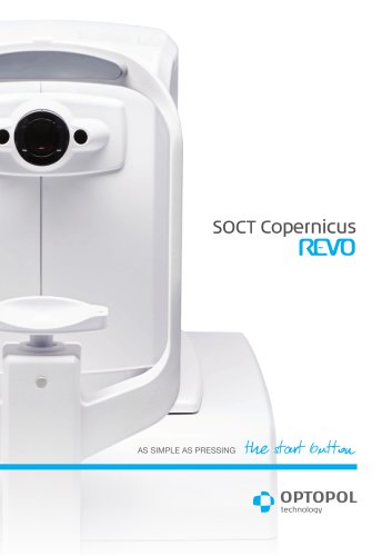 SOCT Coperncius REVO
6 Pages
 REVO FC
6 Pages
 REVO NX
12 Pages

- Analysis software
- Radiology software
- Hospital software
- Optopol fixed ophthalmic examination
- Automated software
- Traceability software
- Capture software
- Software module
- AI-assisted software
- Artificial intelligence software
- Import software
- 3D software
- Ophthalmoscope
- Screening software
- Analysis software module
- Medical software module
- Retinal camera
- Medical imaging software module
- Management software module
- Ophthalmic software