- Catalogs
- Optopol Technology
- REVO FC
REVO FC
1 /6Pages

REVO FC
1 /6Pages
Catalog excerpts

Our supreme experience in Spectral Domain OCT technology allows us to provide you with a single versatile device featuring high resolution OCT and true colour fundus imaging for time and space efficiency. SOCT with the complete Fundus Camera functionality The combination of an All in One OCT technology with a Full Colour Fundus Camera in one compact system gives you high quality OCT images and a detailed colour image for a multipurpose diagnosis. Simultaneous capture of color fundus images of eye diseases and OCT examinations in a single shot saves time and space. Now you can use the REVO FC...
Open the catalog to page 2
FUNDUS CAMERA What makes the REVO FC truly unique is its integrated non-mydriatic 12.3 Mpix Fundus Camera into all in one OCT device capable of capturing detailed colour images of ultra-high quality. The REVO FC is fully automated, safe and easy to use. The advanced optical system ensures high quality imaging at 45° viewing angle. Color Fundus image capture is possible with a pupil as small as 3.3 mm. For OCT 2.4 mm is enough. Easy to use Fundus image processing tools deliver a stunning retinal image. Available modes allow to see detailed photos of a single or both eyes as well as a time comparison...
Open the catalog to page 3
A single 3D macula scan performs both Retina and Ganglion cells analysis. The software automatically recognises 8 retinal layers which assists with a precise diagnosis and the mapping of any changes in the patient’s condition. A variety of result analysis and presentation methods allows for the best selection suitable to increase efficiency of work. Progression Morphology Progression Quantification Revo’s standard high density scanning capability and blood vessel structure recognition enable a precise alignment of past and current scans Operator can analyze changes in morphology, quantified progression...
Open the catalog to page 4
WIDEFIELD SCAN 12x12 mm Widefield Central scan is perfect for fast and precise screening of the patient’s retina. Peripheral scanning reveals diseases in far periphery. Combined view of two examinations of peripheral scan 12mm + 12 mm. Done in external software. T-OCT™ is a pioneering way to provide detailed corneal Curvature maps by using a posterior dedicated OCT. Anterior, Posterior surface and Corneal Thickness allow to provide the True Net Curvature information. With Net power, the precise understading of the patient’s corneal condition comes easily and is free of errors associated with...
Open the catalog to page 5
FUNDUS CAMERA Type Non-mydriatic fundus camera Photography type Color Min. pupil size for fundus 3.3 mm OPTICAL COHERENCE TOMOGRAPHY Technology Spectral Domain OCT Light source SLED Bandwidth 50 nm half bandwidth Scanning speed 80 000 measurements per second Axial resolution 5 pm in tissue, 2.6 pm digital Transverse resolution 12 pm, typical 18 pm Overall scan depth 2.4 mm Focus adjustment range -25 D to +25 D Scan range Posterior 5 mm to 12 mm, Angio 3 mm to 9 mm, Anterior 3 mm to 16 mm Scan types 3D, Angio1, Radial (HD), B-scan (HD), Raster (HD), Cross (HD), TOPO, AL Fundus alignment IR, Live...
Open the catalog to page 6All Optopol Technology catalogs and technical brochures
 REVO NX 130
16 Pages
 REVO 80
8 Pages
 REVO 60
8 Pages
 REVO FC 130
16 Pages
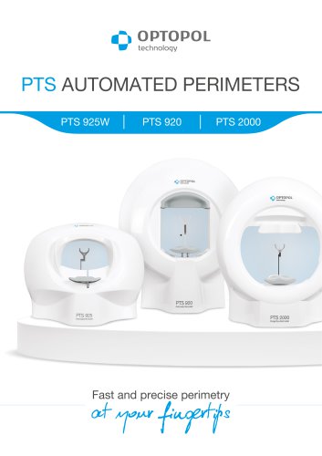 PTS AUTOMATED PERIMETERS
8 Pages
Archived catalogs
 revo-fc130
16 Pages
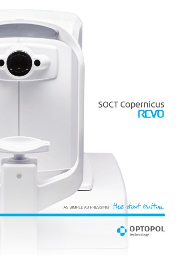 SOCT Coperncius REVO
6 Pages
 REVO NX
12 Pages

- Analysis software
- Radiology software
- Hospital software
- Automated software
- Fixed ophthalmic examination
- Capture software
- Traceability software
- Software module
- AI-assisted software
- Artificial intelligence software
- Import software
- 3D software
- Ophthalmoscope
- Screening software
- Medical software module
- Analysis software module
- Retinal camera
- Ophthalmic software
- Medical imaging software module
- Management software module