
- Catalogs
- Optopol Technology
- REVO FC

REVO FC
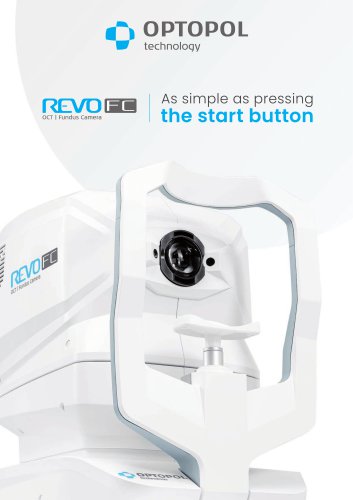
REVO FC
- Ease of Use: Simplifies examinations with a single start button and voice-guided instructions, reducing patient chair time.
- Compact Design: Small footprint and single cable connection make it ideal for small exam rooms.
- Multi-functionality: Functions as a color fundus camera, OCT imaging device, and biometry tool.
- High-Quality Imaging: Equipped with a 12.3 Mpix non-mydriatic fundus camera and advanced optical systems.
- Advanced Software: Features AI Denoise, real-time eye tracking, and comprehensive analysis tools for retina and glaucoma.
- Extended Depth Imaging: Provides deeper scans for challenging cases, suitable for highly myopic patients.
- AI Retina Segmentation: Utilizes AI for accurate retinal layer recognition.
- Ultra-Widefield Module: Offers up to 105° imaging for comprehensive peripheral visualization.
- Single Page Report: Integrates OCT and visual field results for glaucoma management.
- Anterior Segment Imaging: Detailed imaging of anterior eye structures without additional attachments.
- Glaucoma Analysis: Tools for nerve fiber layer and optic nerve head analysis.
- Widefield Analysis: Rapid assessment of retina and glaucoma with comprehensive data reports.
- Cataract Mode: Enhances visualization beneath opaque layers.
- Keratoconus Screening: Provides corneal curvature maps and classification tools.
Catalog excerpts
Assimpleas pressing
Open the catalog to page 1
lution continues New OCT standard Multiple Functions in One Device Once again the REVO FC goes beyond the limits of standard OCT. With its new software, the REVO FC enables a full functionality from the cornea to the retina, combining the potential of several devices. With just a single REVO FC device, you can measure, quantify, calculate, and track changes from the cornea to the retina over time. OCT easier than ever Simply position the patient and press the START button to acquire examinations of both eyes. The REVO FC guides the patient through the process with vocal messages, which increases...
Open the catalog to page 2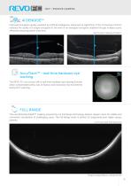
AI DENOISETM Improved tomogram quality powered by Artificial Intelligence. Advanced AI algorithms of the AI Denoise function enhance the quality of a single tomogram to the level of an averaged tomogram obtained through multiple scans, effectively reducing patient chair time. Raw Tomogram / AiDenoise Tomogram AccuTrackTM – real-time hardware eye tracking The REVO FC now comes with a real-time hardware eye tracking function which compensates blinks, loss of fixation, and involuntary eye movements during OCT scanning. FULL RANGE The new Extended Depth™ imaging, powered by our Full Range technology,...
Open the catalog to page 3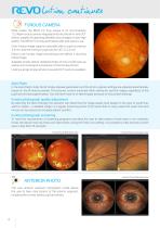
lution continues FUNDUS CAMERA What makes the REVO FC truly unique is its non-mydriatic 12.3 Mpix fundus camera integrated into into this all-in-one OCT device, capable of capturing detailed colour images of ultra-high quality. The REVO FC is fully automated, safe, and easy to use. Color fundus image capture is possible with a pupil as small as 3.3 mm, and the minimum pupil size for OCT is 2.4 mm. Easy to use fundus image processing tools deliver a stunning retinal image. Available modes deliver detailed photos of one or both eyes as well as a chronological comparison of the fundus photos. Linking...
Open the catalog to page 4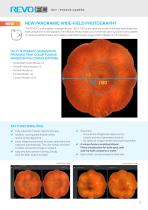
REVONEW PANORAMIC WIDE-FIELD PHOTOGRAPHY The REVO FC now enables coverage of up to 100° (~150° at the spherical center of the eye), providing a new wide perspective in retinography. In the Mosaic Photo mode, up to 9 internal fixation positions are available to capture peripheral areas and create a wide-field mosaic image. Patient dilation is not mandatory. FULLY AUTOMATIC ACQUISITION PROVIDES TRUE-COLOR FUNDUS IMAGES IN FIVE CONFIGURATIONS: • Horizontal Fundus Mosaic x3 • Vertical Fundus Mosaic x3 V Fully automatic mosaic capture process V Intuitive, voice-guided fixation shifts speed up the...
Open the catalog to page 5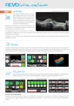
Automatically detected 10 retinal layers The new layer segmentation for the posterior segment is based on artificial intelligence, resulting in more accurate recognition of retinal layer boundaries. The AI system enhances the accuracy of clinical evaluation and supports more reliable assessment of retinal pathology. High level of detection accuracy empowers the clinician with more detailed screening, increasing the effectiveness of everyday eye care. AI segmentation will be important for follow-up examinations, enabling a more accurate diagnosis when analysing pathology over time. It can also...
Open the catalog to page 6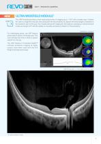
Ultra-Widefield Module1 The UWF module provides a new wide perspective of imaging up to ~105° with a single scan. It allows the user to image the macular area along with the far periphery to capture the early stages of disease in the posterior part of the eye. The module allows 3D imaging for full analysis, averaging in enhancement mode, and angio OCT with the ability to visualise perfusion problems in the periphery. Widefield 21 3D report For challanging cases,, the UWF feature will provide an option of a Radial scan. This scan will take less time to attain a proper signal from the eye. The...
Open the catalog to page 7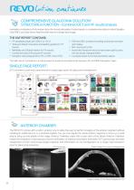
lution continues COMPREHENSIVE GLAUCOMA SOLUTION1 STRUCTURE & FUNCTION - Combined OCT and VF results analysis 1 Invaluable combination of information about the functional quality of vision based on comprehensive data on retinal Ganglion Cells, RNFL and Optic Nerve Head for both eyes on a single report page. The S&F report contains: • VF sensitivity results (24-2/30-2 or 10-2) • Total and Pattern Deviation probability graphs for VF results • Reliability and Global indices for VF results • Combined map of Structure & Function • Ganglion cells analysis (GCL+IPL or NFL+GCL+IPL) • ONH and NFL analysis...
Open the catalog to page 8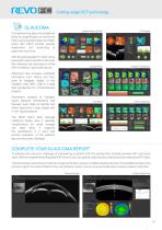
GLAUCOMA Comprehensive glaucoma analytical tools for quantification of the Nerve Fiber Layer, Ganglion layer and Optic Head with DDLS provide precise diagnostics and monitoring of glaucoma over time. With the gold standard 14 optic nerve parameters and a new Rim to Disc and Rim Absence, the description of the ONH condition is quick and precise. Ganglion Both Ganglion Progression Advanced view provides combined information from Retina and Disc scan to integrate details of the Ganglion cells, RNFL, ONH in a wide field perspective for comprehensive analysis. Asymmetry Analysis of Ganglion layers...
Open the catalog to page 9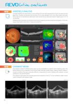
Widefield analysis A single Widefield 3D examination is now sufficient for the rapid assessment of both the retina and the glaucoma. Visualize and assess the thickness of the retina, ganglion cell, nerve fibers layers and optic nerve head on comprehensive data report when performing a rapid examination mapping up to 15×15 mm section. Widefield report presents horizontal and vertical tomograms and will include the topography of the disc, supporting helpful observation of glaucoma patients. 3D Widefield scan Cataract mode The cataract mode in the REVO series opens up new possibilities for patients...
Open the catalog to page 10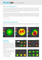
OCT TOPOGRAPHY1,2 T-OCT™ is a pioneering way to provide detailed corneal Curvature maps by using posterior dedicated OCT. Parameters of the anterior and posterior surfaces, and the corneal thickness provide True Net Curvature information. With the Net power, a precise understanding of the patient’s corneal condition is easy and free of errors associated with modelling of posterior surface of the cornea. The REVO T-OCT module provides axial maps, tangential mas, total Power map, height maps, epithelium and corneal thickness maps. The corneal topography module clearly shows the changes in the cornea...
Open the catalog to page 11All Optopol Technology catalogs and technical brochures
 REVO NX 130
16 Pages
 REVO 80
8 Pages
 REVO 60
8 Pages
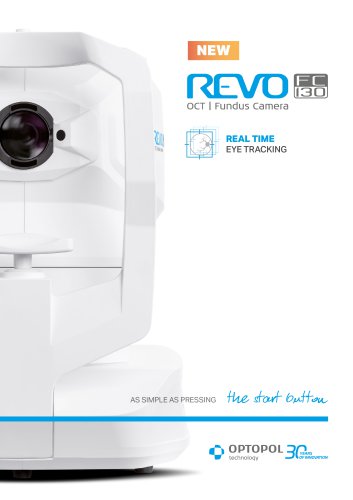 REVO FC 130
16 Pages
Archived catalogs
 PTS AUTOMATED PERIMETERS
8 Pages
 revo-fc130
16 Pages
 SOCT Coperncius REVO
6 Pages
 REVO FC
6 Pages
 REVO NX
12 Pages

- Analysis software
- Radiology software
- Hospital software
- Automated software
- Optopol fixed ophthalmic examination
- Traceability software
- Software module
- Capture software
- AI-assisted software
- Artificial intelligence software
- Import software
- 3D software
- Ophthalmoscope
- Screening software
- Medical software module
- Analysis software module
- Retinal camera
- Medical imaging software module
- Ophthalmic software
- Management software module