
Catalog excerpts

lution continues Optopol engineering team, the designers of the first commercially available Spectral Domain OCT in the world, are proud to present the latest innovation, the world`s fi rst B-OCT and T-OCT for standard posterior OCT. Our supreme experience in Spectral Domain OCT allows us to provide the market with a state of the art instrument which comes with new advanced technologies and remarkable simplicity of operation. The latest software release sets up new demands for daily OCT routine in a modern ophthalmic practice. The new modules expand the diagnostic range of OCT by the addition...
Open the catalog to page 2
offers the latest standards available in OCT technology Single A single 3D Retina scan performs both Retina and Glaucoma analyses. The software automatically recognizes 8 retinal layers which assists with a precise diagnosis and the mapping of any changes in the patient’s condition. A variety of result analysis and presentation methods allows for the best selection suitable to increase efficiency of work Progression Morphology Progression Quantif cation Revo’s standard high density scanning capability and blood vessel structure recognition enable a precise alignment of past and current...
Open the catalog to page 3
lution continues ANGIOGRAPHY SOCT¹ This non-invasive dye free technique allows the visualization of the microvasculature of the retina. Both blood flow and structural visualiza-tion give additional diagnostic information about many retinal diseases. Angiography scan allows assessment of the structural vasculature of the macula, the periphery or the optic disc. Extremely short scanning times of 1,6 seconds in standard resolution or 3 seconds in high resolution. Now Angiography OCT can become a routine in your diagnostic practice. QUANTIFICATION The quantification tool provides a...
Open the catalog to page 4
offers the latest standards available in OCT technology MOST COMPLETE SET OF ANGIO OCT ANALYSIS VIEWS Standard Single View Detailed Single View Software allows to observe, track and compare changes in the microvasculature of the retina in both eyes. ANGIOGRAPHY MOSAIC1 The Angiography mosaic delivers high-detail images over a large field of the retina. Available modes allow to see predefined region of the retina in a convenient way. Manual mode allows to scan the d e s i re d re g i o n . B u i l t - i n a n a l y t i c s allow to see vascular layers, enface or thick-ness maps. Healthy...
Open the catalog to page 5
lution continues ANTERIOR For a standard anterior examination, an additional lens or attachment is not required. This allows the examiner to quickly complete the scanning procedure. Cornea Single Presentation of the results for both eyes allows quick and precise evaluation of the condition of the patient’s anterior segment. Epithelium and Pachymetry map are included in the standard package. An additional adapter included in the package increases the range of clinical application in Anterior chamber observation. OCT Gonioscopy* Wide cornea scan, Descemet’s membrane detachment (DMD) and...
Open the catalog to page 6
offers the latest standards available in OCT technology TOPOGRAPHY OCT¹ T-OCT™ is a pioneering way to provide detailed corneal Curvature maps by using posterior dedicated OCT. Anterior, Posterior surfaces and Corneal Thickness allow to provide the True Net Curvature information. With Net power, the precise understading of the patient’s corneal condition comes easily and is free of errors associated with modelling of posterior surface of the cornea. SOCT T-OCT module provides Axial maps, Tangential maps, Total Power map, Height maps, Epithelium and Corneal thickness maps. Corneal topography...
Open the catalog to page 7
GLAUCOMA Comprehensive glaucoma analytical tools for quantification of the Nerve Fiber Layer, Ganglion layer and Optic Head with DDLS allow for the precise diagnosis and monitoring of glaucoma over time. With the golden standard 14 optic nerve parameters and a new Rim to Disc and Rim Absence the description of ONH condition is quick and precise. Advanced view which provides combined information from Retina and Disc scan to integrate details of the Ganglion cells, RNFL, ONH in a wide field perspective for comprehensive analysis for both eyes. Asymmetry Analysis of Ganglion layers between...
Open the catalog to page 8
offers the latest standards available in OCT technology COMPREHENSIVE GLAUCOMA SOLUTION STRUCTURE & FUNCTION - Combined OCT and VF results analysis Invaluable combination of information about the functional quality of vision with comprehensive data on retinal Ganglion Cells, RNFL and Optic Nerve Head for both eyes on a single report page. The S&F report contains the following: • VF sensitivity results (24-2/30-2 or 10-2) • Total and Pattern Deviation probability graphs for VF results • Reliability and Global indices for VF results • Combined map of Structure & Function • Ganglion cell...
Open the catalog to page 9
offers the latest standards available in OCT technology BIOMETRY OCT¹ Single view Result review B-OCT® Innovative method of using the posterior OCT device to measure ocular structure along eye axis. OCT Biometry provides a complete set of Biom-etry parameters: Axial Length AL, Central Cornea Thickness CCT, Anterior Chamber Depth ACD, Lens Thickness LT. VERIFY YOUR MEASURMENT VISUALLY All measurement callipers are shown on all boundaries of OCT image provided by REVO. Now, you can visually verify, identify and if need be, make corrections as to which structure of the eye has been measured....
Open the catalog to page 10
Central 12 mm scan, Enhance Mode to provide vitreous and choroid details. Cornea scan, Posterior graft (DSAEK) detachment Anterior scan, Cornea Guttata with corneal scaring * Images courtesy of Prof. Edward Wylegała MD, PhD * Images courtesy of Prof. Edward Wylegała MD, PhD *Images courtesy of Bartosz L. Sikorski MD, PhD
Open the catalog to page 11
Light source: Scanning speed: Axial resolution: Transverse resolution: Overall scan depth: Minimum pupil size: Focus adjustment range: Scan range: Posterior 5 mm to 12 mm, Angio 3 mm to 9 mm, Anterior 3 mm to 16 mm Scan types: 3D, Angio¹, Radial (HD), B-scan (HD), Raster (HD), Cross (HD), TOPO, AL, ACD Fundus image: Live Fundus Reconstruction Alignment method: Fully automatic, Automatic, Manual Retina analysis: Retina thickness, Inner Retinal thickness, Outer Retinal thickness RNFL+GCL+IPL thickness, GCL+IPL thickness, RNFL thickness, RPE deformation, IS/OS thickness Angiography OCT¹:...
Open the catalog to page 12All Optopol Technology catalogs and technical brochures
-
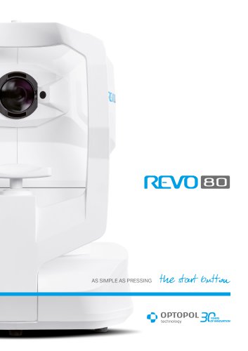 REVO 80
8 Pages
-
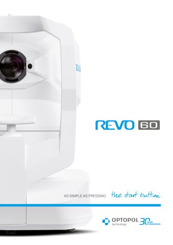 REVO 60
8 Pages
-
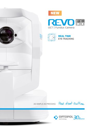 REVO FC 130
16 Pages
-
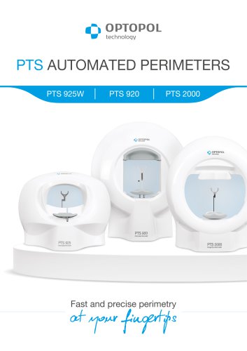 PTS AUTOMATED PERIMETERS
8 Pages
-
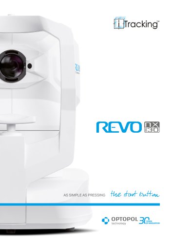 REVO NX 130
12 Pages
Archived catalogs
-
 revo-fc130
16 Pages
-
 REVO NX
12 Pages
-
 REVO FC
6 Pages
-
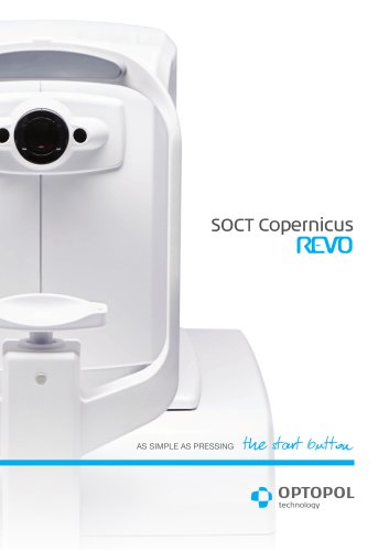 SOCT Coperncius REVO
6 Pages
