- Catalogs
- Optopol Technology
- REVO NX 130
REVO NX 130
1 /16Pages
REVO NX 130
1 /16Pages
Catalog excerpts

20 Years of REVO lution
Open the catalog to page 1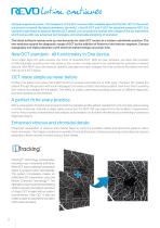
lution continues Optopol engineering team, the designers of the first commercially available Spectral Domain OCT in the world, are proud to present the latest innovation, the world`s first B-OCT and T-OCT for standard posterior OCT. Our supreme experience in Spectral Domain OCT allows us to provide the market with a state of the art instrument which comes with new advanced technologies and remarkable simplicity of operation. The latest software release sets up new demands for daily OCT routine in a modern ophthalmic practice. The new modules expand the diagnostic range of OCT by the addition...
Open the catalog to page 2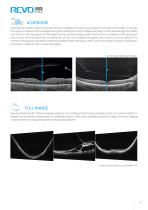
AI DENOISE Improved tomogram quality powered by Artificial Intelligence. Advanced AI algorithms enhance the quality of a single tomogram to the level of an averaged tomogram obtained through multiple scanning. The AI DeNoise algorithm filters out noise form the tomogram for the highest and smoothest image quality. The function is available on all tomograms and in every tab featuring them, including the 3D tab. On averaged tomograms the function is on by default. The moment a tomogram is loaded for review the software starts denosing it. After a short moment the original “undenoised” tomogram...
Open the catalog to page 3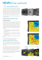
lution continues ANGIOGRAPHY SOCT1 This non-invasive dye free technique allows the visualization of the microvasculature of the retina. Both blood flow and structural visualization give additional diagnostic information about many retinal diseases. Angiography scan allows assessment of the structural vasculature of the macula, the periphery or the optic disc. Extremely short scanning times of 1,6 seconds in standard resolution or 3 seconds in high resolution. Now Angiography OCT can become a routine in your diagnostic practice. ANGIO ANALYSIS METHODS QUANTIFICATION The quantification tool provides...
Open the catalog to page 4
Maximum Intesity Projection – the MIP algorithm Choose better visualization of angio data for analysis with the Maximum Intensity Projection (MIP) feature. This tool is useful for visualizing OCT-A data as it enables easier identification and tracking of high-intensity structures such as blood vessels. AIP A COMPLETE SET OF ANGIO OCT ANALYSIS VIEWS Software allows to observe, track and compare changes in the microvasculature of the retina in both eyes. Standard Single View Detailed Single View
Open the catalog to page 5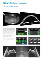
lution continues ANGIOGRAPHY MOSAIC1 The Angiography mosaic delivers high-detail images over a large field of the retina. Available modes allow to see predefined region of the retina in a convenient way. In manual mode it is possible to scan the desired region. Built-in analytics allow the user to see vascular layers, enface or thickness maps. Healthy patient, Angio Mosaic mode: 7×7 mm PDR, Angio Mosaic mode: 10x10 mm *Images courtesy of Bartosz L. Sikorski MD, PhD an optional software module to purchas
Open the catalog to page 6
ANTERIOR CHAMBER Built-in anterior lens allows the user to perform the imaging of the anterior segment without installing additional lens or forehead adapter. Now you can display the whole anterior segment or focus on a small area to bring out the details of the image. Cornea Single New Anterior Chamber protocols with a fast view of the whole Anterior Chamber. Now you can evaluate gonioscopy situation and verify cataract lens easier and faster. Presentation of the results for both eyes allows quick and precise evaluation of the condition of the patient’s anterior segment. Epithelium and Pachymetry...
Open the catalog to page 7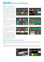
lution continues TOPOGRAPHY OCT1 T-OCT™ is a pioneering way to provide detailed corneal Curvature maps by using posterior dedicated OCT. Ante-rior, Posterior surfaces and Corneal Thickness provide the True Net Curvature information. With the Net power a precise understanding of the patient’s corneal condition comes easily and is free of errors associated with model-ling of posterior surface of the cornea. SOCT T-OCT module provides Axial maps, Tangential mas, Total Power map, Height maps, Epithelium and Corneal thickness maps. Corneal topography module clearly shows the changes in the cornea...
Open the catalog to page 8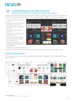
Widefield analysis A single Widefield 3D examination is now sufficient for the rapid assessment of both the retina and the glaucoma. Visualize and assess the thickness of the retina, ganglion cell, nerve fibers layers and optic nerve head on comprehensive data report when performing a rapid examination mapping up to 15×15 mm section. Widefield report presents horizontal and vertical tomograms and will include the topography of the disc creating helpful observation of glaucoma patients. Cataract mode The cataract mode in the REVO series opens up new possibilities for patients with challening cases....
Open the catalog to page 9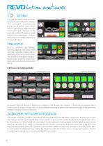
lution continues GLAUCOMA Comprehensive glaucoma analytical tools for quantification of the Nerve Fiber Layer, Ganglion layer and Optic Head with DDLS enable the user to perform precise diagnosis and monitoring of glaucoma over time. Ganglion Both Ganglion Progression With the golden standard 14 optic nerve parameters and a new Rim to Disc and Rim Absence the description of ONH condition is quick and precise. Advanced view which provides combined information from Retina and Disc scan to integrate details of the Ganglion cells, RNFL, ONH in a wide field perspective for comprehensive analysis. With...
Open the catalog to page 10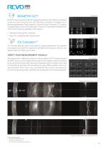
REVOCOMPREHENSIVE GLAUCOMA SOLUTION STRUCTURE & FUNCTION - Combined OCT and VF results analysis Invaluable combination of information about the functional quality of vision with comprehensive data on retinal Ganglion Cells, RNFL and Optic Nerve Head for both eyes on a single report page. The S&F report contains the following: • Total and Pattern Deviation proba bility graphs for VF results • Reliability and Global indices for VF results • Combined map of Structure & Function • Ganglion cells analysis (GCL+IPL or NFL+GCL+IPL) • ONH and NFL analysis including charts and comparison tables • Nasal...
Open the catalog to page 11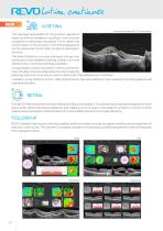
AI Retina Automatically detected 10 retinal layers This new layer segmentation for the posterior segment is based on artificial intelligence, resulting in more accurate recognition of retinal layer boundaries. The AI system has a direct impact on the accuracy of the clinical assessment and the assessment of the status of areas of pathology in the retina. This level of detection accuracy empowers the eye care and results in more detailed screening. Overall, it is a more effective way of running a pathology evaluation. AI segmentation will be important for follow-up examinations, bringing a more...
Open the catalog to page 12All Optopol Technology catalogs and technical brochures
 REVO 80
8 Pages
 REVO 60
8 Pages
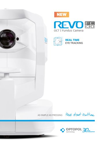 REVO FC 130
16 Pages
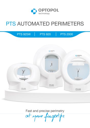 PTS AUTOMATED PERIMETERS
8 Pages
Archived catalogs
 revo-fc130
16 Pages
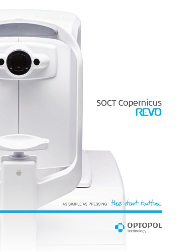 SOCT Coperncius REVO
6 Pages
 REVO FC
6 Pages
 REVO NX
12 Pages

- Analysis software
- Radiology software
- Hospital software
- Automated software
- Fixed ophthalmic examination
- Capture software
- Traceability software
- AI-assisted software
- Artificial intelligence software
- Import software
- Ophthalmoscope
- 3D software
- Screening software
- Analysis software module
- Medical software module
- Retinal camera
- Medical imaging software module
- Ophthalmic software
- Management software module