- Catalogs
- Optopol Technology
- REVO NX
REVO NX
1 /12Pages

REVO NX
1 /12Pages
Catalog excerpts
lution continues Optopol engineering team, the designers of the first commercially available Spectral Domain OCT in the world, are proud to present the latest innovation, the world`s first B-OCT and T-OCT for standard posterior OCT. Our supreme experience in Spectral Domain OCT allows us to provide the market with a state of the art instrument which comes with new advanced technologies and remarkable simplicity of operation. The latest software release sets up new demands for daily OCT routine in a modern ophthalmic practice. The new modules expand the diagnostic range of OCT by the addition...
Open the catalog to page 2
offers the latest standards available in OCT technology RETINA A single 3D Retina scan performs both Retina and Glaucoma analyses. The software automatically recognizes 8 retinal layers which assists with a precise diagnosis and the mapping of any changes in the patient’s condition. A variety of result analysis and presentation methods allows for the best selection suitable to increase efficiency of work. Progression Morphology Progression Quantification Revo’s standard high density scanning capability and blood vessel structure recognition enable a precise alignment of past and current scans....
Open the catalog to page 3
lution continues ANGIOGRAPHY SOCT1 This non-invasive dye free technique allows the visualization of the microvasculature of the retina. Both blood flow and structural visualization give additional diagnostic information about many retinal diseases. Angiography scan allows assessment of the structural vasculature of the macula, the periphery or the optic disc. Extremely short scanning times of 1,6 seconds in standard resolution or 3 seconds in high resolution. Now Angiography OCT can become a routine in your diagnostic practice. MOST COMPLETE SET OF VIEWS Standard Single View Detailed Single View...
Open the catalog to page 4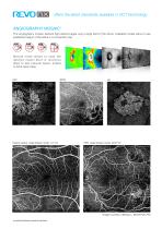
offers the latest standards available in OCT technology ANGIOGRAPHY MOSAIC1 The Angiography mosaic delivers high-detail images over a large field of the retina. Available modes allow to see predefined region of the retina in a convenient way. Manual mode allows to scan the d e s i re d re g i o n . B u i l t - i n a n a l y t i c s allow to see vascular layers, enface or thick-ness maps. Healthy patient, Angio Mosaic mode: 7×7 mm PDR, Angio Mosaic mode: 10x10 mm *Images courtesy of Bartosz L. Sikorski MD, PhD 1 an optional software module to purchas
Open the catalog to page 5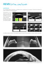
lution continues ANTERIOR For a standard anterior examination, an additional lens or attachment is not required. This allows the examiner to quickly complete the scanning procedure. Cornea Single Angle Both P re s e n t a t i o n o f t h e results for both eyes allows quick and precise evaluation of the condition of the patient’s anterior segment. Epithelium and Pachymetry map are included in the standard package. An additional adapter included in the package increases the range of clinical application in Anterior chamber observation. Angle to Angle scan, narrow angles Wide cornea scan, Descemet’s...
Open the catalog to page 6
offers the latest standards available in OCT technology TOPOGRAPHY OCT1 T-OCT™ is a pioneering way to provide detailed corneal Curvature maps by using posterior dedicated OCT. Anterior, Posterior surface and Corneal Thickness allow to provide the True Net Curvature information. With Net power, the precise understading of the patient’s corneal condition comes easily and is free of errors associated with modelling of posterior surface of the cornea. SOCT T-OCT module provides Axial maps, Tangential maps, Total Power map, Height maps, Epithelium and Corneal thickness maps. Corneal topography module...
Open the catalog to page 7
lution continues GLAUCOMA Comprehensive glaucoma analytical tools for quantification of the Nerve Fiber Layer, Ganglion layer and Optic Head with DDLS allow for the precise diagnosis and monitoring of glaucoma over time. With the golden standard 14 optic nerve parameters and a new Rim to Disc and Rim Absence the description of ONH condition is quick and precise. Advance Retina & ONH Ganglion Both Ganglion Progression Advanced view which provides combined information from Retina and Disc scan to integrate details of the Ganglion cells, RNFL, ONH in a wide field perspective for comprehensive analysis....
Open the catalog to page 8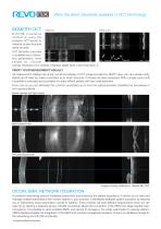
offers the latest standards available in OCT technology Single view Result review B-OCT® Innovative method of using the posterior OCT device to measure ocular structure along eye axis. OCT Biometry provides a complete set of Biometry parameters: Axial Length AL, Central Cornea Thickness CCT, Anterior Chamber Depth ACD, Lens Thickness LT. VERIFY YOUR MEASURMENT VISUALLY All measurement callipers are shown on all boundaries of OCT image provided by REVO. Now, you can visually verify, identify and if need be, make corrections as to which structure of the eye has been measured. With a simple cursor...
Open the catalog to page 9
CLINICAL IMAGES Central 12 mm scan Choroidal observation Sample of angio Manual mode *Images courtesy of Bartosz L. Sikorski MD, PhD
Open the catalog to page 10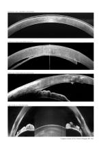
Cornea scan, Fuchs’ Endothelial Corneal Dystrophy Cornea scan, Posterior graft (DSAEK) detachment Anterior scan, Cornea Guttata with corneal scaring Angle to Angle scan, Ciliary Body visible *Images courtesy of Prof. Edward Wylęgała MD, Ph
Open the catalog to page 11
0 197 www.optopol.com Local Distributor: OPTOPOL Technology sp. z o.o. ul. Zabia 42, 42-400 Zawiercie, Poland Tel/Fax: +48 32 6709173 S [email protected]
Open the catalog to page 12All Optopol Technology catalogs and technical brochures
 REVO NX 130
16 Pages
 REVO 80
8 Pages
 REVO 60
8 Pages
 REVO FC 130
16 Pages
 PTS AUTOMATED PERIMETERS
8 Pages
Archived catalogs
 revo-fc130
16 Pages
 SOCT Coperncius REVO
6 Pages
 REVO FC
6 Pages

- Analysis software
- Radiology software
- Hospital software
- Automated software
- Fixed ophthalmic examination
- Capture software
- Traceability software
- AI-assisted software
- Artificial intelligence software
- Import software
- 3D software
- Ophthalmoscope
- Screening software
- Medical software module
- Analysis software module
- Retinal camera
- Ophthalmic software
- Medical imaging software module
- Management software module