- Catalogs
- Optopol Technology
- SOCT Coperncius REVO
SOCT Coperncius REVO
SOCT Coperncius REVO
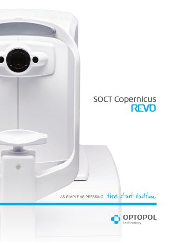
The SOCT Copernicus REVO is a modern Spectral Domain Optical Coherence Tomography (OCT) device designed for simplicity and efficiency in eye examinations. It is suitable for various clinical settings due to its compact design and advanced diagnostic capabilities.
Ease of Use
The device simplifies the examination process by allowing operators to position the patient and press the start button to conduct scans of both eyes. Vocal guidance enhances patient comfort and reduces chair time. Customizable scanning protocols improve workflow efficiency.
Design and Installation
The SOCT Copernicus REVO features a small footprint, flexible positioning options, and a single-cable connection, making it ideal for small examination rooms. It functions effectively as both a screening and advanced diagnostic tool.
Image Quality
Noise reduction technology ensures high-quality OCT images, crucial for early disease detection. The device offers a widefield scan of 12x12 mm for comprehensive retinal screening, including peripheral areas.
Diagnostic Capabilities
- Retina: A single 3D macula scan provides both retina and glaucoma analysis, automatically recognizing eight retinal layers for precise diagnosis.
- Glaucoma: Comprehensive tools for analyzing nerve fiber layers and optic nerve head, with asymmetry analysis for early detection.
- Angiography: Non-invasive, dye-free visualization of retinal microvasculature, aiding in the diagnosis of retinal diseases.
- Anterior Segment: Standard examinations require no additional lens, with an adapter available for expanded clinical applications.
Technical Specifications
The device operates with a spectral domain OCT technology, featuring a scanning speed of 27,000 measurements per second and an axial resolution of 5 µm. It supports various scan programs and offers detailed analysis for both retina and glaucoma conditions.
Additional Features
The device includes a fixation target with an OLED display, adjustable focus range, and operates on a power supply of 100–240 V. It is compact and lightweight, weighing 23 kg.
Conclusion
The SOCT Copernicus REVO is a versatile and efficient OCT device, offering advanced diagnostic capabilities and ease of use, making it a valuable addition to any modern ophthalmic practice.
Catalog excerpts
SOCT Copernicus
Open the catalog to page 1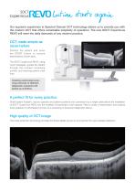
lution starts again Our supreme experience in Spectral Domain OCT technology allows us to provide you with the modern OCT that offers remarkable simplicity of operation. The new SOCT Copernicus REVO will meet the daily demands of any modern practice. OCT made simple as never before Position the patient and press the START button to acquire examinations of both eyes. The SOCT Copernicus REVO, using vocal messages, guides the patient through the process increasing comfort and reducing patient chair time. Creating customised scanning protocols of different diagnostic scenarios will speed up workflow....
Open the catalog to page 2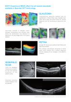
SOCT Copernicus REVO offers the all newest standards available in Spectral OCT technology. GLAUCOMA Comprehensive glaucoma analysis tools for quantification of Nerve Fiber Layer, Ganglion layer Optic Nerve Head with DDLS allows for precise diagnosis and the monitoring of glaucoma over time. Asymmetry Analysis of Ganglion layers between hemispheres and between eyes allows the identification and detection of glaucoma in its early stages and in non-typical patients. RETINA A single 3D macula scan performs both Retina and Glaucoma analysis. The software automatically recognises 8 retinal layers which...
Open the catalog to page 3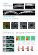
ANTERIOR For a standard anterior examination, no additional lens is required. This allows the examiner to quickly complete the scanning procedure. Additional adapter provided with the device increases range of clinical application in Anterior chamber observation. ANGIOGRAPHY SOCT* This non-invasive dye free technique allows the visualisation of the microvasculature of the retina. Both blood flow and structural visualisation will give additional information in the diagnosis of many retinal diseases. Now Angiography SOCT can be a routine diagnosis information in your practice. FOLLOW UP Revo’s...
Open the catalog to page 4
• Choroidal observation • Wide Central scan • Sclera and Anterior Structure
Open the catalog to page 5
Light source Scanning speed Axial resolution Transverse resolution Overall scan depth Scan range Scan program 3D, Radial, B-scan, Raster, Cross Fundus image Live Fundus Reconstruction Alignment method Fully automatic, Automatic Retina analysis Retina thickness, Inner retinal thickness, Outer retinal thickness, RNFL+GCL+IPL thickness, GCL+IPL thickness, RNFL thickness, RPE deformation, IS/OS thickness Glaucoma analysis RNFL, ONH morphology, DDLS, Ganglion analysis as RNFL+GCL+IP and GCL+IPL, OU and Hemisphere asymmetry Anterior Pachymetry map, Epithelium map, LASIK flap assesment, Angle Assessment,...
Open the catalog to page 6All Optopol Technology catalogs and technical brochures
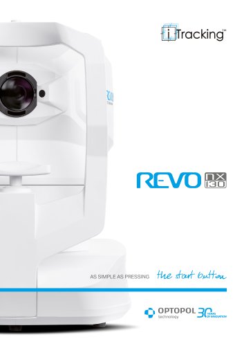 REVO NX 130
16 Pages
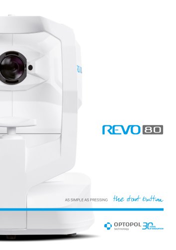 REVO 80
8 Pages
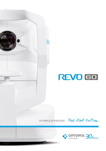 REVO 60
8 Pages
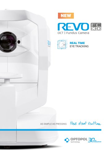 REVO FC 130
16 Pages
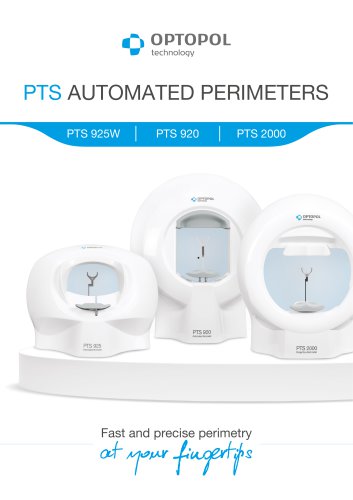 PTS AUTOMATED PERIMETERS
8 Pages
Archived catalogs
 revo-fc130
16 Pages
 REVO FC
6 Pages
 REVO NX
12 Pages

- Analysis software
- Radiology software
- Hospital software
- Automated software
- Fixed ophthalmic examination
- Capture software
- Traceability software
- Software module
- AI-assisted software
- Artificial intelligence software
- Import software
- Ophthalmoscope
- 3D software
- Screening software
- Analysis software module
- Medical software module
- Retinal camera
- Medical imaging software module
- Ophthalmic software
- Management software module