- Catalogs
- OrthoPediatrics
- PediLoc ® Extension Osteotomy
PediLoc ® Extension Osteotomy

PediLoc ® Extension Osteotomy
The PediLoc® Extension Osteotomy Plate is part of the OrthoPediatrics® system, designed for use in children and adolescents to fit the anatomy of the distal femur. It is available in left and right configurations with sizes of 6, 8, and 10 hole plates. The plates are pre-contoured, low profile, and periosteal sparing to minimize soft tissue damage.
Indications
The plate is indicated for opening and closing wedge osteotomies, fractures, mal-unions, and non-unions of the femur.
Background
This technique addresses severe knee flexion contractures, often seen in children with cerebral palsy. The procedure may involve hamstring lengthening and patellar tendon adjustments. Careful consideration is given to avoid excessive valgus and manage neurovascular tension.
Surgical Technique
Preparation: Determine the desired femoral extension using clinical examination and x-rays. Position the patient supine and prepare the surgical site.
Patient Positioning: Ensure visualization of the hip, knee, and ankle joints. Apply a sterile tourniquet if possible.
Surgical Approach: Use fluoroscopy to mark the distal femoral physis and perform a lateral approach. Expose the distal femur subperiosteally.
Position Implant: Use a guide wire to align the plate, ensuring all screw holes engage bone. Adjust as necessary to prevent excess valgus.
Provisional Plate Fixation: Align the plate with the tibia and insert distal locking screws for provisional fixation. Mark the osteotomy level and remove the plate.
Perform Osteotomy: Use an oscillating saw to cut the femur and remove a bone wedge. Consider femoral shortening to manage neurovascular tension.
Attach Plate: Align the plate and secure it with locking screws distally and proximally. Adjust for coronal alignment and contour as needed.
Important Notes
The device is supplied non-sterile and is intended for single use. It is not approved for spinal fixation. The technique is a guideline and not a substitute for professional medical judgment.
Catalog excerpts

PediLoc® Extension Osteotomy Plate (PLEO) Left PLEO Plates Sizes: 6, 8 and 10 hole plates Right PLEO Plates Sizes: 6, 8 and 10 hole plates
Open the catalog to page 1
PediLoc® Extension Osteotomy Plate The technique description herein is made available to the healthcare professional to illustrate the author’s suggested treatment for the uncomplicated procedure. In the final analysis, the preferred treatment is that which addresses the needs of the specific patient. PediLoc® Extension Osteotomy Plating System Surgical Technique Introduction PediLoc® Extension Osteotomy Plate . . . . . . . . . . . . . . .2 Indications . . . . . . . . . . . . . . . . . . . . . . . . . . . . . . . . . . . . . . .2 Background . . . . . . . . . . . . . . . . . . . . . . . . . ....
Open the catalog to page 2
Severe knee flexion contractures in children remain a difficult treatment problem. This technique will be used primarily for patients with persistent knee flexion contracture during the stance phase of gait, most of whom have cerebral palsy. Residual static knee flexion contracture is treated by a compensatory osteotomy of the distal femur of the exact same degree as the knee flexion contracture. • In many cases this procedure is either preceded, or accompanied by, a hamstring lengthening. • In most cases the procedure should be accompanied by a patellar tendon shortening or alternatively, a...
Open the catalog to page 3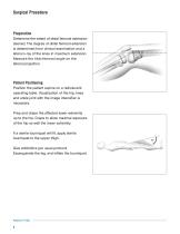
Surgical Procedure Preparation Determine the extent of distal femoral extension desired. The degree of distal femoral extension is determined from clinical examination and a lateral x-ray of the knee in maximum extension. Measure the tibio-femoral angle on the lateral projection. Patient Positioning Position the patient supine on a radiolucent operating table. Visualization of the hip, knee and ankle joint with the image intensifier is necessary. Prep and drape the affected lower extremity up to the hip. Drape to allow maximal exposure of the hip as well the lower extremity. If a sterile tourniquet...
Open the catalog to page 4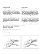
Surgical Approach Position Implant Using fluoroscopy, mark out the level of the distal femoral physis. Make a standard lateral approach to the distal femur If planning a concomitant patellar tendon shortening then curve the distal portion of the incision anteriorly over the patellar tendon. Place a guide wire (2mm k-wire) parallel to and just proximal to the distal femoral physis, slightly anterior to the midpoint of the femur. This will help later with translation. Slide the plate over the guide wire to ensure that all the screw holes in the distal flared portion of the plate will engage bone....
Open the catalog to page 5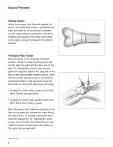
Surgical Procedure Position Implant Using fluoroscopy, hold the plate against the shaft of the distal lateral femur. Sometimes the plate will need to be contoured to prevent excess valgus following osteotomy. Utilize the threaded drill guides in the distal holes while contouring to prevent damage to the internal threads. Provisional Plate Fixation Place the knee in the maximally extended position. Using the distal targeting guide with handle, align the plate with the long axis of the tibia. Or, alternatively, use the plate as your guide and align the plate to the long axis of the tibia. In the...
Open the catalog to page 6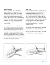
Perform Osteotomy Attach Plate Using an oscillating saw, cut the femur perpendicular to the long axis of the femur. Make a second cut to match the angle of correction desired and remove the wedge of bone. Once the osteotomy is completed, perform a trial reduction. Pay careful attention to the tension created posteriorly and also to translation. The distal fragment should be translated posteriorly to minimize the anterior bump. At this point the femora-tibial angle should be 0 degrees with the former flexion angle now shifted to the osteotomy site. Once trial reduction looks satisfactory, apply...
Open the catalog to page 7
PediLoc® Extension Osteotomy to be used in conjunction with PediLoc® Small Fragment (3.5mm) and PediLoc® Large Fragment (4.5mm). CAUTION: Federal law restricts this device to sale by or on the order of a Physician. CAUTION: Devices are supplied Non-Sterile. Clean and sterilize before use according to instructions. CAUTION: Implant components are single-use. Do not reuse. CAUTION: This device is not approved for screw attachment or fixation to the posterior elements (pedicles) of the cervical, thoracic or lumbar spine. This document is intended exclusively for experts in the field, i.e. physicians...
Open the catalog to page 8All OrthoPediatrics catalogs and technical brochures
 PediFrag ™ System 2.7/3.5
30 Pages
 PediFlex
24 Pages
 PediLoc
2 Pages
 Cannulated Screw
8 Pages
- Bone plate
- Compression plate
- Metallic compression plate
- Locking compression plate
- Rectangular table
- Distal compression plate
- Orthopedic surgery instrument kit
- Compression bone screw
- Metallic compression bone screw
- Proximal compression plate
- Arthrodesis nail
- Lateral compression plate
- Medial compression plate
- Tibia compression plate
- General purpose compression bone screw
- External fixation system
- Metallic intramedullary nail
- Cannulated compression bone screw
- Arthrodesis plate
- Proximal fixation intramedullary nail