

- Products
- Catalogs
- News & Trends
- Exhibitions
i-scan Atlas for Gastroenterology

i-scan Atlas for Gastroenterology
The document discusses i-scan and i-scan Optical Enhancement (OE) technologies, which improve gastrointestinal endoscopy by enhancing tissue and vascular visualization. These technologies are crucial for diagnosing and planning treatments for various gastrointestinal conditions.
i-scan technology, including Surface Enhancement (SE) and Tone Enhancement (TE), aids in lesion detection and characterization. Optical Enhancement (OE) provides detailed vascular pattern visualization, helping differentiate neoplastic from non-neoplastic tissues.
Specific settings for i-scan and i-scan OE modes are outlined, enhancing image quality and supporting clinical decision-making.
- Oesophagus: i-scan and i-scan OE assist in diagnosing early squamous cell carcinoma, Barrett’s oesophagus, and oesophageal squamous cell cancer by visualizing vascular changes.
- Stomach: These technologies assess gastric lesions and diagnose conditions like Gastrointestinal Stromal Tumor (GIST) and ECL cell micronodular hyperplasia.
- Duodenum: i-scan technologies help diagnose Crohn’s disease and characterize ampullary lesions.
- Colon: i-scan and i-scan OE analyze colonic polyps, conduct surveillance in familial adenomatous polyposis, and detect serrated polyps.
The document concludes with information on i-scan certification and ongoing studies validating the technology's clinical effectiveness.
i-scan and i-scan OE technologies significantly advance endoscopic imaging, providing detailed visualization that supports accurate diagnosis and treatment planning in gastroenterology.
- Barrett’s Oesophagus: Enhanced imaging techniques like i-scan and OE are used for surveillance and detection of neoplasia.
- Gastric Lesions: i-scan and OE enhance surface and vascular patterns, aiding in early gastric dysplastic lesion detection.
- Gastrointestinal Stromal Tumor (GIST): These technologies visualize submucosal masses and confirm diagnoses with endoscopic ultrasound and elastography.
- Hyperplastic Polyps: i-scan and OE aid in diagnosing and characterizing hyperplastic polyps.
- Crohn’s Disease: Advanced imaging detects subtle mucosal lesions and assesses inflammatory conditions.
Experts highlight the effectiveness of i-scan and i-scan OE in enhancing mucosal and vascular patterns, aiding early cancer detection, and improving diagnostic accuracy.
- i-scan 1 (SE): Used for initial lesion detection with enhanced surface resolution.
- i-scan 2 (TE): Characterizes vascularity and microvascular patterns.
- i-scan 3 (OE): Provides detailed visualization of inflammatory appearances and vascular patterns.
- Crohn’s Disease Diagnosis: i-scan techniques identify fibrinous erosions, leading to Crohn’s disease diagnosis.
- Ampullary Lesion Characterization: i-scan imaging determines lesion extension and grade, guiding treatment.
- Colonic Polyp Analysis: i-scan and OE assess diminutive polyps, enhancing optical biopsy accuracy.
- Surveillance in Familial Adenomatous Polyposis: i-scan techniques detect and characterize lesions, supporting therapeutic decisions.
A 70-year-old male in a CRC screening program had lesions detected in the colon, examined using i-scan technology, revealing a Kudo type 3L pit pattern, indicating a tubular adenoma with low-grade dysplasia. The lesion was resected using endoscopic mucosal resection (EMR).
i-scan technology, including OE, enhances mucosal and vascular pattern visualization during endoscopy, aiding in gastrointestinal pathology detection and diagnosis.
Studies demonstrate i-scan technology's efficacy in improving gastrointestinal lesion detection and characterization, enhancing diagnostic yield in conditions like Barrett's esophagus and IBD.
The i-scan certification program includes training modules, videos, and webinars to ensure proficiency in using the technology for enhanced diagnostic accuracy.
Catalog excerpts

i-scan Atlas for Gastroenterology Courtesy of Dr Federico Buffoli, Ospedale di Cremona, Italy Case studies from clinical practice with HD+, i-scan and i-scan OE
Open the catalog to page 1
PEWTAXMEDICAL Index Visible excellence. Gastrointestinal endoscopy with HD+, i-scan and i-scan OE. Introduction: Visible excellence 3 A leap forward for in vivo histology 4 Proven i-scan settings 4 i-scan and i-scan OE, a unique combination of digital and optical enhancements to support the clinical pathway 5 Case Reports - Diagnosis of early squamous cell carcinoma with i-scan, i-scan OE - Diagnosis of a Barrett's oesophagus with i-scan, i-scan OE and MagniView 10 - Diagnosis of an oesophageal squamous cell cancer with i-scan, i-scan OE - Detection and resection of an early neoplastic lesion...
Open the catalog to page 2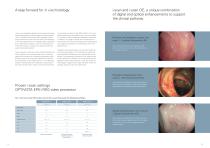
i-scan is an established digital enhancement technology supporting detection, characterization and demarcation. i-scan 1 Surface Enhancement (SE) highlights surface tissue architecture which may be used for the detection of circumscribed lesions or to highlight diffuse alteration (e.g. inflammation, atrophy). i-scan 2 or Tone Enhancement (TE) focuses on vascular and tissue architecture, supporting characterization. The introduction of the new i-scan Optical Enhancement (OE) in addition to the existing i-scan family, will bring additional information on mucosal and especially vascularization patterns...
Open the catalog to page 3
The i-scan Leaders The i-scan task force is a community of highly experienced doctors who have used i-scan in their clinical practice for many years. PENTAX Medical has been working closely with this group so that their best practices and scientific evidence in i-scan can be shared in a peer environment. Its members participate in training and case discussion forums and ultimately contribute to the highest quality of care to patients. Professor Dr. Ralf Kiesslich HSK Wiesbaden, Germany Professor Dr. Pradeep Bhandari Queen Alexandra Hospital, UK Dr. Silvia Sanduleanu Maastricht UMC+, The Netherlands...
Open the catalog to page 4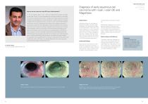
How do you use i-scan and i-scan OE in your clinical practice? “In my own practice I rely on i-scan 1 (SE) as my default initial mode for interrogat- Diagnosis of early squamous cell carcinoma with i-scan, i-scan OE and MagniView OESOPHAGUS STO M AC H DUODENUM COLON ing the upper GI tract. This has superseded the white light mode for me and helps me to assess and detect early cancers. The depth and image resolution is excellent at defining mucosal patterns that when disordered or irregular lead one to suspect neoplasia. My own preference is then to switch to i-scan 2 (TE) to better characterize...
Open the catalog to page 5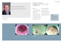
Diagnosis of a Barrett´s oesophagus with i-scan, i-scan OE and MagniView OESOPHAGUS STO M AC H DUODENUM COLON How do you use i-scan and i-scan OE in your clinical practice? “I use systematically advanced imaging techniques such as i-scan and i-scan OE in the work-up of patients with suspicion of Barrett’s associated neoplasia to help in the detection of small subtle lesions.“ Patient history Patient outcome and follow-up A 57 year old male patient with history of reflux disease was referred after surveillance endoscopy with random biopsies detected high grade dysplasia. The patient was scheduled...
Open the catalog to page 6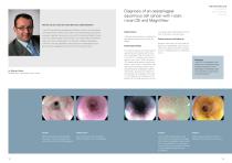
OESOPHAGUS STO M AC H DUODENUM COLON Diagnosis of an oesophageal squamous cell cancer with i-scan, i-scan OE and MagniView How do you use i-scan and i-scan OE in your clinical practice? “i-scan OE combines the versatility and flexibility of i-scan with even sharper and crisper pictures. While i-scan 1 (SE) is my standard setting during every procedure, because it gives you the extra bit of contrast and detail to detect even very subtle changes of the mucosa, i-scan 3 (OE) offers an extremely sharp view of the surface pattern, for example in Barrett’s oesophagus.. Patient history A 60 year old...
Open the catalog to page 7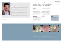
How do you use i-scan and i-scan OE in your clinical practice? “In my own practice I rely on i-scan 1 (SE) as my default initial mode for interrogat- Detection and resection of an early neoplastic lesion in a Barret’s oesophagus with i-scan, i-scan OE and MagniView OESOPHAGUS STO M AC H DUODENUM COLON ing the upper GI tract. This has superseded the white light mode for me and helps me to assess and detect early cancers. The depth and image resolution is excellent at defining mucosal patterns that when disordered or irregular lead one to suspect neoplasia. My own preference is then to switch to...
Open the catalog to page 8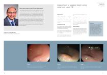
How do you use i-scan and i-scan OE in your clinical practice? Assessment of a gastric lesion using i-scan and i-scan OE O E S O P H AG U S STOMACH DUODENUM COLON ”The PENTAX Medical High-Definition White Light (HD WL) image is a major advance but when I switch i-scan 1 (SE) on then the image becomes sharper and crisper without losing any other attribute so I don’t see any reason why i-scan 1 (SE) should not be left on at all times. Once I find a subtle abnormality or any obvious lesion then I switch on i-scan 2 (TE) and that really highlights the lesion from the surrounding normal mucosa. Once...
Open the catalog to page 9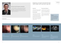
O E S O P H AG U S STOMACH DUODENUM COLON Diagnosis of a Gastrointestinal Stromal Tumor (GIST) with i-scan and i-scan OE How do you use i-scan and i-scan OE in your clinical practice? “I usually use as default i-scan 1 (SE) as a detection mode and I usually turn on i-scan 3 (OE) when I want to better visualize the mucosal and vascular pattern. I find it particularly useful in Barrett’s oesophagus to diagnose high grade dysplasia area, for rectal and colonic polyp to decide between ESD or piece meal EMR when i-scan 3 (OE) shows aspect of carcinoma in situ or limited to sm1“ Patient outcome and...
Open the catalog to page 10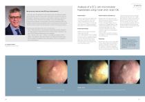
How do you use i-scan and i-scan OE in your clinical practice? “I think that i-scan OE represents a further step forward in the diagnostic capabilities of endoscopy. With the i-scan system we can see the mucosa and the lesions with different visualizations that allows to collect different information, specific for each enhance. We can choose the most suitable i-scan function depending on: anatomical site, pathology or just according to the way the endoscopist feels more confident to be able to make the diagnosis. After my first experience with i-scan OE I feel that it allows to better study the...
Open the catalog to page 11All Pentax catalogs and technical brochures
 the IMAGINA i10c Endoscopes
2 Pages
 INSPIRA™
2 Pages
 PENTAX Medical i20c
2 Pages
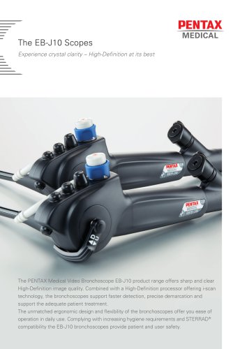 The EB-J10 Scopes
2 Pages
Archived catalogs
 i-scan Mini-Atlas
17 Pages
 Confocal Endomicroscopy
2 Pages
 More than meets the eye
16 Pages
 NaviAid™ BGE
4 Pages
 Video Gastroscopes
2 Pages
 Monochrome Gastroscopes
2 Pages
 Fiber Gastroscopes
2 Pages
 ENT Video Scope
2 Pages
 SAFE-3000
4 Pages
 EB-1575K
4 Pages
 Monochrome Bronchoscopes
2 Pages
 Video Bronchoscopes
2 Pages
 video endoscopy for ENT
2 Pages
 Intubation
2 Pages
 CLASSIC-CART
2 Pages
 90i and 90K
2 Pages
 EC-3490Ti
2 Pages
 PENTAX Medical Brochure
5 Pages
 The i10 endoscope series
2 Pages
 HD+ Bronchoscope EB-1990i
2 Pages
 Speech, Voice & Swallowing
8 Pages
 Digital Video Processor
2 Pages
 EPK-i and EPK-i5000
6 Pages
 HD+ and i-scan
8 Pages
 NaviAid AB
2 Pages
 90i-90k
2 Pages


- Endoscopy forceps
- Electrosurgical forceps
- Bipolar forceps
- LED light source
- Medical video endoscope
- Endoscope light source
- Biopsy forceps
- High-definition medical video endoscope
- Compact light source
- Endoscopy video processor
- Video bronchoscope
- Hemostatic forceps
- HD imaging video processor
- Video gastroscope
- 4K imaging video processor
- Video processor with USB port
- High-definition video bronchoscope
- Pre-cleaning station
- Video colonoscope
- 3D imaging video processor