- Products
- Catalogs
- News & Trends
- Exhibitions
i-scan Mini-Atlas

i-scan Mini-Atlas
The document provides an overview of the advanced imaging capabilities of HD+ and i-scan technology in gastrointestinal endoscopy, emphasizing their role in enhancing mucosal surface visualization and aiding in the detection and characterization of abnormalities.
HD+ endoscopy, combined with i-scan technology, improves visualization of mucosal surfaces by highlighting vascular details and tissue architecture. The i-scan modes, Surface Enhancement (SE) and Tone Enhancement (TE), assist in detecting and characterizing lesions.
- Barrett’s Oesophagus: Utilization of HD+ and i-scan facilitated in vivo characterization and targeted biopsies, leading to accurate diagnosis and treatment.
- Familial Adenomatous Polyposis (FAP): i-scan SE and TE improved lesion delineation and characterization, aiding in the management of duodenal adenomas.
- Gastric Adenoma: HD+ and i-scan allowed for clear visualization and successful endoscopic submucosal dissection of a gastric adenoma.
- Gastric High-Grade Dysplasia: i-scan enhanced detection of dysplastic features, supporting targeted biopsies and effective treatment.
- Gastric Polyp Analysis: i-scan provided detailed characterization of mucosal patterns and vascularization, guiding treatment decisions.
- Chronic Diarrhoea and Adenomatous Lesion: i-scan SE and TE helped demarcate a sessile lesion in the transverse colon, identified as a tubular adenoma with low-grade dysplasia.
- Ulcerative Colitis and Dysplasia: i-scan SE and TE characterized a large lesion in the right colon, leading to piecemeal mucosa resection and confirmation of low-grade dysplasia.
- Colorectal Cancer Detection: i-scan techniques highlighted a large neoplasia in the ascending colon, leading to a right hemicolectomy and diagnosis of well-differentiated adenocarcinoma.
- Non-Polypoid Colorectal Neoplasms: i-scan SE and TE characterized a non-polypoid lesion in the ascending colon as an adenomatous lesion, removed via EMR.
- Colonic LST Lesion: i-scan SE and TE indicated a high risk of invasive cancer in a large superficial lesion, leading to en-bloc resection and diagnosis of tubulovillous adenoma with high-grade dysplasia.
- Pedunculated Polyp Analysis: i-scan confirmed a pedunculated polyp in the sigmoid colon as a tubular adenoma, removed via snare polypectomy.
- Post-Polypectomy Scar Examination: i-scan examination revealed residual adenomatous tissue, leading to transanal endoscopic microsurgery.
- Sessile Polyp Characterization: i-scan characterized a sessile polyp in the rectum, leading to successful resection and confirmation as a tubular adenoma.
The document highlights the clinical value of HD+ and i-scan technologies in enhancing endoscopic diagnosis and treatment of gastrointestinal and colorectal lesions, contributing to improved patient outcomes through enhanced diagnostic accuracy and therapeutic interventions.
Catalog excerpts

i-scan Mini-Atlas Case studies from clinical practice with HD + and i-scan.
Open the catalog to page 1
Visible excellence. Gastrointestinal endoscopy with HD and i-scan. + Index Introduction3 HD and i-scan at a glance + 4 5 – PENTAX i-scan in characterization of Barrett’s oesophagus 6 7 – PENTAX i-scan in surveillance endoscopy in a patient with familial adenomatous polyposis 8 9 – PENTAX i-scan in diagnosis of gastric adenoma 10 11 – Gastric high-grade dysplasia of the antrum demonstrated with PENTAX i-scan 12 13 – Analysis of a gastric polyp using PENTAX i-scan 14 15 – Detection and characterization of colorectal polyps using PENTAX i-scan 16 – 17 Detection and characterization...
Open the catalog to page 2
Your eyes are familiar with natural, sharp vision. Why give them anything less? Advantages of HD and i-scan at a glance. + HD in tubulo-villous adenoma + • Supports fast orientation and detection • Significant improvement in the visibility and evaluation of minute lesions • ntegrated zoom function for more detailed I inspection of suspicious surface structures Enrich your endoscopic options with HD and i-scan. + The i-scan technology is based on the post processing of reflected light. i-scan can be switched on and off by simply pushing a button on the endoscope. The different i-scan modes...
Open the catalog to page 3
PENTAX i-scan in characterization of Barrett’s oesophagus. Patient history A 67-year-old man with reflux symptoms was referred for an upper gastrointestinal endoscopy after no relief with proton pump inhibitors. At endoscopy, he was found to have a Barrett’s oesophagus C3M5, according to the Prague classification. Endoscopic findings With HD + White Light endoscopy, we identified an area of mucosa that appeared slightly featureless on initial examination. With the addition of i-scan SE, this area became more evident. Normal glandular mucosa surrounded a central area where the mucosal pattern...
Open the catalog to page 4
PENTAX i-scan in surveillance e ndoscopy in a patient with familial adenomatous polyposis. Patient history A 42-year-old woman with familial adenomatous polyposis (FAP) syndrome presented for esophagogastroduodenoscopy. She underwent her first colonoscopy five years ago, because both her father and uncle died of colorectal cancer at the ages of 45 and 50, respectively. Her first colonoscopy revealed more than 100 small polyps throughout the colon which were all tubular adenomas. DNA analysis showed a mutation of the APC gene confirming the diagnosis of FAP. A protective total colectomy was...
Open the catalog to page 5
PENTAX i-scan in diagnosis of gastric adenoma. Patient history A 55-year-old female patient was referred to our GI unit for endoscopic submucosal dissection of a lesion at the gastric angular fold. Biopsies taken at the referring hospital revealed an adenoma with high-grade intraepithelial neoplasia (HGIN). Endoscopic findings The upper gastrointestinal endoscopy showed a flat-depressed lesion (IIa c – according to the Paris classification) at the angular fold measuring approximately 42 mm x 37 mm in diameter. HD + White Light imaging clearly shows the lesion. By adding i-scan SE, the margins...
Open the catalog to page 6
Gastric high-grade dysplasia of the antrum demonstrated with PENTAX i-scan. Patient history This 62-year-old male patient was brought in with a long history of intermittent upper abdominal pain and a known gastric ulcer of at least 30 years. He was found to have an incidental positive faecal occult blood test and underwent colonoscopy and upper gastrointestinal endoscopy. There was no history of melaena or vomiting. Apart from hypertension, for which he was taking anti-hypertensives, he was well. Endoscopic findings The oesophagus and duodenum were normal. The stomach distended easily and views...
Open the catalog to page 7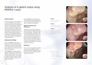
Analysis of a gastric polyp using PENTAX i-scan. Patient history A 56-year-old patient was admitted to her local emergency room with malaena and was found to be significantly anaemic. After aggressive resuscita tion, she underwent endoscopy and was found to have a pre-pyloric ulcer which was bleeding. She underwent endotherapy with adrenaline and endoclips and was discharged. Biopsies from the ulcer edge taken at the initial examination showed high-grade dysplasia. She returned 4 weeks later for a repeat endoscopy. Endoscopic findings At the repeat HD + White Light endoscopy, an obvious polyp...
Open the catalog to page 8
Detection and characterization of colorectal polyps using PENTAX i-scan. Patient history A 61-year-old male patient at the outpatient clinic had chronic diarrhoea with four loose stools a day. His medical history showed type 2 diabetes and COPD. A colonoscopy was performed to rule out colon pathology as a cause for his symptoms. Endoscopic findings HD + White Light colonoscopy (image 1) revealed a sessile lesion (Paris 0 Is) – located in the transverse colon and measuring approx. 30 mm x 20 mm. Optical examination using i-scan SE and TE (images 2 and 3) allowed better demarcation of the...
Open the catalog to page 9
Detection and characterization of dysplasia in ulcerative colitis using PENTAX i-scan. Patient history A 79-year-old man with long-standing ulcerative colitis, currently in remission, was referred to our endoscopy unit for surveillance colonoscopy. Endoscopic findings The rectum and left colon showed a relatively normal pit pattern and some pseudopolyps, indicating chronic inflammation. Using HD + White Light image, a 60 mm large circumscribed lesion with irregular margins (0 IIa LST of non-granular – type) was found in the right colon. i-scan SE and TE helped to demarcate the borders or...
Open the catalog to page 10
Analysis of colorectal cancer using PENTAX i-scan. Patient history A 72-year-old man was referred to our emergency department because of dizziness and discomfort. On physical examination, the skin appeared pale. Laboratory investigation showed anaemia with haemoglobin of 7.8 g/dl (reference 13 17), serum iron of 35 µg/dl – (40 160) and C-reactive protein of – 90 mg/l (< 5). Faecal occult blood testing was positive. EGD was normal. Endoscopic findings HD + White Light colonoscopy (image 1) revealed a large neoplasia in the ascending colon with a central depression. Analysis of the remaining...
Open the catalog to page 11All Pentax catalogs and technical brochures
 the IMAGINA i10c Endoscopes
2 Pages
 INSPIRA™
2 Pages
 PENTAX Medical i20c
2 Pages
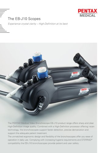 The EB-J10 Scopes
2 Pages

Archived catalogs
 Confocal Endomicroscopy
2 Pages
 More than meets the eye
16 Pages
 NaviAid™ BGE
4 Pages
 Video Gastroscopes
2 Pages
 Monochrome Gastroscopes
2 Pages
 Fiber Gastroscopes
2 Pages
 ENT Video Scope
2 Pages
 SAFE-3000
4 Pages
 EB-1575K
4 Pages
 Monochrome Bronchoscopes
2 Pages
 Video Bronchoscopes
2 Pages
 video endoscopy for ENT
2 Pages
 Intubation
2 Pages
 CLASSIC-CART
2 Pages
 90i and 90K
2 Pages
 EC-3490Ti
2 Pages
 PENTAX Medical Brochure
5 Pages
 The i10 endoscope series
2 Pages
 HD+ Bronchoscope EB-1990i
2 Pages
 Speech, Voice & Swallowing
8 Pages
 Digital Video Processor
2 Pages
 EPK-i and EPK-i5000
6 Pages
 HD+ and i-scan
8 Pages
 NaviAid AB
2 Pages
 90i-90k
2 Pages


- Endoscopy forceps
- Electrosurgical forceps
- Bipolar forceps
- Medical video endoscope
- Biopsy forceps
- High-definition medical video endoscope
- Endoscopy video processor
- Endoscopy column
- Video bronchoscope
- Hemostatic forceps
- HD imaging video processor
- Video gastroscope
- 4K imaging video processor
- Video processor with USB port
- Pre-cleaning station
- High-definition video bronchoscope
- Video colonoscope
- High-definition video gastroscope
- 3D imaging video processor
- Endoscope pre-cleaning station