- Products
- Catalogs
- News & Trends
- Exhibitions
More than meets the eye

More than meets the eye
Catalog excerpts
Video Endomicroscopy PENTAX Europe GmbH LIFE CARE Julius-Vosseler-Straße 104 22527 Hamburg Germany Tel.: +49 40 / 5 61 92 - 0 Fax: +49 40 / 5 60 42 13 E-Mail: [email protected] PENTAX Italia S.r.l. LIFE CARE Via Dione Cassio, 15 20138 Milano Italy Tel.: +039 / 02 50 99 58 1 Fax: +039 / 02 50 99 58 60 E-mail: [email protected] PENTAX U.K. Limited LIFE CARE Pentax House Heron Drive, Langley Slough SL3 8PN United Kingdom Tel.: +44 17 53 / 79 27 23 Fax: +44 17 53 / 79 27 94 E-Mail: [email protected] PENTAX France S.A.S. LIFE CARE 112 quai de Bezons P. B. 204 95106 Argenteuil France Tel.: +33 1 / 30 25 75 75 Fax: +33 1 / 30 25 75 76 E-Mail: [email protected] PENTAX Nederland B.V. LIFE CARE Lage Mosten 35 4822 NK Breda Netherlands Tel.: +31 76 / 5 31 30 31 Fax: +31 76 / 5 31 30 00 E-Mail: [email protected] MTM/01/09/05/00000/02 HOYA Corporation PENTAX Tokyo office 2-36-9, Maeno-cho Itabashi-Ku 174-8639 Tokyo Japan Tel.: +81 33 / 9 60 51 55 Fax: +81 35 / 3 92 67 24 More than meets the eye Mini-Atlas of Confocal Laser Endomicroscopy
Open the catalog to page 1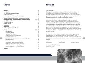
Index Preface Introduction The confocal laser endoscope Contrast agents Procedure of confocal laser endoscopy Preface 3 4–5 6–7 8 9 – 10 Colorectal Cancer: Screening with endomicroscopy Ulcerative colitis: Surveillance with endomicroscopy Barrett‘s oesophagus Helicobacter pylori Gastric Cancer Conclusion References Confocal pattern classification 2 11 12 – 13 14 15 16 17 18 19 Cases Normal colon Colorectal Cancer Colorectal adenomas Inflammation in ulcerative colitis Barrett’s oesophagus Advanced adenocarcinoma of the distal oesophagus Normal stomach Gastric Cancer Helicobacter pylori associated...
Open the catalog to page 2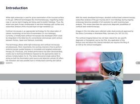
Introduction White-light endoscopy is used for gross examination of the mucosal surface in the gut. Different technologies like chromoendoscopy, magnifying endoscopy or narrow band imaging are being used to predict histology. Thus, the dream and goal of every endoscopist is real-time histology with surface and subsurface analysis of the mucosal layer at high resolution [1]. Confocal microscopy is an appropriate technology for the observation of cellular morphology at the time of examination (in vivo histology). Currently, a miniaturised confocal microscope was developed which could be integrated...
Open the catalog to page 3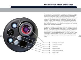
The confocal laser endoscope The components of the confocal laser endoscope are based on the integration of a confocal laser microscope in the distal tip of a conventional video endoscope, which enables confocal microscopy in addition to standard video endoscopy. The diameter of both the distal tip and the insertion tube is 12.8 mm. The distal tip contains an air and water jet nozzle, two light guides, an auxiliary water jet channel (used for topical application of the contrast agent) and a 2.8 mm working channel. Actuation of imaging plane depth relative to the surface of the tissue is controlled...
Open the catalog to page 4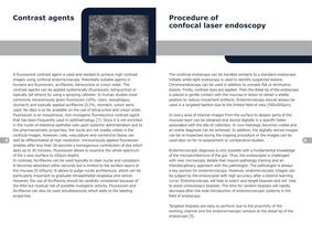
Contrast agents 8 Procedure of confocal laser endoscopy A fluorescent contrast agent is used and needed to achieve high contrast images using confocal endomicroscopy. Potentially suitable agents in humans are fluorescein, acriflavine, tetracycline or cresyl violet. The contrast agents can be applied systemically (fluorescein, tetracycline) or topically (all others) by using a spraying catheter. In human studies most commonly intravenously given fluorescein (10%; colon, oesophagus, stomach) and topically applied acriflavine (0.2%; stomach, colon) were used. No data is so far available on the use...
Open the catalog to page 5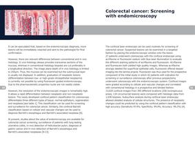
Colorectal cancer: Screening with endomicroscopy It can be speculated that, based on the endomicroscopic diagnosis, more lesions will be immediately resected and sent to the pathologist for final confirmation. However, there are relevant differences between conventional and in vivo histology. In vivo histology always provides transverse sections of the mucosa, whereas in conventional histology biopsies are cut preferably in a longitudinal direction. The image plane depth of in vivo histology is limited to 250μm. Thus, the mucosa can be examined but the submucosal layer is usually not displayed....
Open the catalog to page 6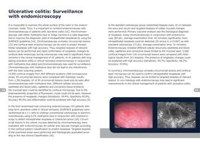
Ulcerative colitis: Surveillance with endomicroscopy 12 It is impossible to examine the whole surface of the colon in the endomicroscopic mode. Thus, it is important to combine endomicroscopy with chromoendoscopy in patients with ulcerative colitis (UC). Panchromoendoscopy with either methylene blue or indigo carmine is a valid diagnostic tool to improve the diagnostic yield of intraepithelial neoplasias by using the SURFACE guidelines [14]. Chromoendoscopy unmasks circumscript lesions [15] and confocal laser endomicroscopy can be used to predict intraepithelial neoplasias with high accuracy....
Open the catalog to page 7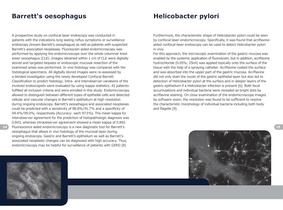
Barrett‘s oesophagus 14 Helicobacter pylori A prospective study on confocal laser endoscopy was conducted in patients with the indications long lasting reflux symptoms or surveillance endoscopy (known Barrett’s oesophagus) as well as patients with suspected Barrett’s associated neoplasias. Fluorescein aided endomicroscopy was performed by applying the endomicroscope over the whole columnar lined lower oesophagus (CLE). Images obtained within 1 cm of CLE were digitally stored and targeted biopsies or endoscopic mucosal resection of the examined areas was performed. In vivo histology was compared...
Open the catalog to page 8
Gastric cancer Confocal laser endomicroscopy was also used to diagnose gastric cancer and precancerous conditions. Endomicroscopy was performed on five ex-vivo gastrectomy specimens and in upper GI endoscopies in vivo in eight patients. Acriflavine hydrochloride dye was used for ex vivo examinations and intravenous fluorescein sodium for in vivo examinations. A standard upper endoscopy was performed, upon which confocal images were obtained at standardized locations in the gastric antrum, body and cardia, before taking biopsy specimens from the same areas for histopathology. Confocal diagnostic...
Open the catalog to page 9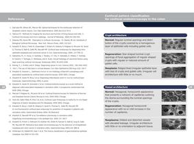
References Confocal pattern classification for confocal endomicroscopy in the colon 1. DaCosta RS, Wilson BC, Marcon NE. Optical techniques for the endoscopic detection of dysplastic colonic lesions. Curr Opin Gastroenterol. 2005 Jan;21(1):70-9. 2. Tadrous PJ. Methods for imaging the structure and function of living tissues and cells: 3. Confocal microscopy and micro-radiology. Journal of Pathology 2000;191:345-354. 3. Delaney PM, Harris MR. Fiberoptics in confocal microscopy. In: Pawley JB, ed. Handbook of Biological Confocal Microscopy. New York: Plenum Press, 1995: 515-523 4. Kiesslich R,...
Open the catalog to page 10All Pentax catalogs and technical brochures
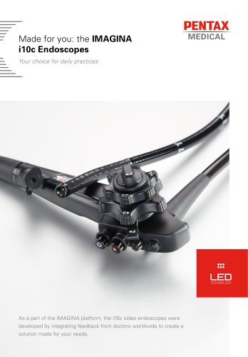 the IMAGINA i10c Endoscopes
2 Pages
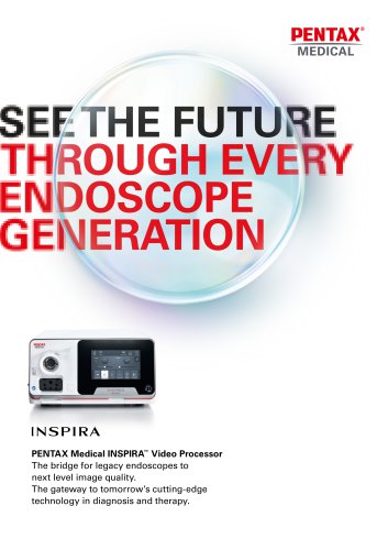 INSPIRA™
2 Pages
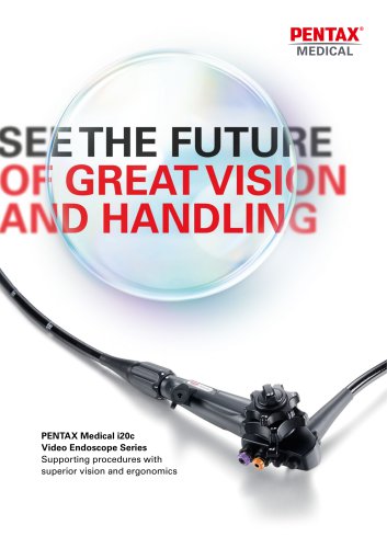 PENTAX Medical i20c
2 Pages
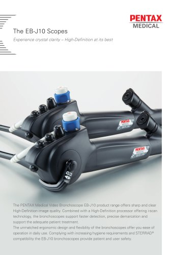 The EB-J10 Scopes
2 Pages
Archived catalogs
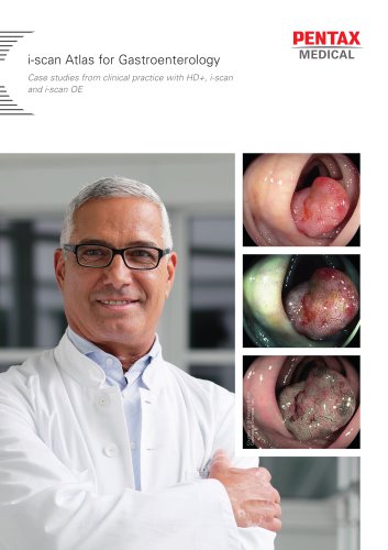
 i-scan Mini-Atlas
17 Pages
 Confocal Endomicroscopy
2 Pages
 NaviAid™ BGE
4 Pages
 Video Gastroscopes
2 Pages
 Monochrome Gastroscopes
2 Pages
 Fiber Gastroscopes
2 Pages
 ENT Video Scope
2 Pages
 SAFE-3000
4 Pages
 EB-1575K
4 Pages
 Monochrome Bronchoscopes
2 Pages
 Video Bronchoscopes
2 Pages
 video endoscopy for ENT
2 Pages
 Intubation
2 Pages
 CLASSIC-CART
2 Pages
 90i and 90K
2 Pages
 EC-3490Ti
2 Pages
 PENTAX Medical Brochure
5 Pages
 The i10 endoscope series
2 Pages
 HD+ Bronchoscope EB-1990i
2 Pages
 Speech, Voice & Swallowing
8 Pages
 Digital Video Processor
2 Pages
 EPK-i and EPK-i5000
6 Pages
 HD+ and i-scan
8 Pages
 NaviAid AB
2 Pages
 90i-90k
2 Pages


- Endoscopy forceps
- Electrosurgical forceps
- Bipolar forceps
- Medical video endoscope
- Biopsy forceps
- High-definition medical video endoscope
- Endoscopy video processor
- Endoscopy column
- Video bronchoscope
- Hemostatic forceps
- HD imaging video processor
- Video gastroscope
- 4K imaging video processor
- Video processor with USB port
- Pre-cleaning station
- High-definition video bronchoscope
- Video colonoscope
- High-definition video gastroscope
- 3D imaging video processor
- Endoscope pre-cleaning station