- Catalogs
- Philips Healthcare
- Philips Ingenuity TF PET/CT system
Philips Ingenuity TF PET/CT system
1 /96Pages

Philips Ingenuity TF PET/CT system
1 /96Pages
Catalog excerpts

Advanced molecular imaging Ingenuity TF PET/CT Clinical case book Oncology cases The print quality of this copy is not an accurate representation of the original.
Open the catalog to page 1
Clinical case book The print quality of this copy is not an accurate representation of the original.
Open the catalog to page 2
Ingenuity TF PET/CT Oncology cases Clinical case book The print quality of this copy is not an accurate representation of the original.
Open the catalog to page 3
Apollo Gleneagles Hospital, 20 Kolkata, India 1. Recurrent fibrolamellar variant of hepatocellular 22 2. Moderately differentiated squamous cell 24 carcinoma of the left lateral border of the tongue, treated by left hemiglossectomy along with left sided modified neck dissection 3. Metastatic adenocarcinoma of right sided 26 cervical lymph node but unknown primary. Treated by right sided radical neck dissection, 2014. Follow up study 4. Metastatic poorly differentiated carcinoma to D8 28 done for unknown primary 5. Adenocarcinoma of the splenic flexure of the 30 colon with infiltration of spleen,...
Open the catalog to page 4
Isala, Zwolle, The Netherlands 54 1. Primary breast cancer with several axillary 56 2. Breast cancer primary with axillary metastases 58 8 mm and very subtle 4 mm parasternal metastatic lesion 3. Mid-esophageal tumor with mediastinal nodes 60 as small as 3-4 mm picked up on FDG PET and degenerative disease in cervical spine 4. Burkitt lymphoma in the lower abdomen 62 mimicking intestinal loops with small upper abdominal mets anterior to liver 5. Pulmonary adenocarcinoma with extensive 64 metastatic spread to mediastinum, axilla, bone and soft tissue 6. Patient with known colon carcinoma 66 7....
Open the catalog to page 5
Images we get from the Ingenuity TF are high quality. They allow me to see small lesions and help to provide the necessary information to my referring physicians and enable more personalized care for their patients. The Ingenuity TF gives me great confidence in my diagnostic interpretations and will serve my purposes now and for years ahead as one of my key instruments as molecular imaging evolves.” Dr. Robert Wagner M.D., M.S.M.I.S., FACR, FACNP Professor, Medical Director, Nuclear Medicine
Open the catalog to page 6
Clinical case book The print quality of this copy is not an accurate representation of the origina
Open the catalog to page 7
Study 1: Innumerable hypermetabolic lesions in the skin, subcutaneous tissue, lungs, liver, kidneys and bones are consistent with metastatic disease. General characteristics Patient Female Scan characteristics 12.9 mCi F18 FDG 62 min uptake time 60 sec/bed Study 2: 10 weeks later. Disease progression seen in the chest, liver and skeleton. General characteristics Patient Age Height Bodyweight Scan characteristics 11.2 mCi F18 FDG 64 min uptake time 60 sec/bed 8 Clinical case book
Open the catalog to page 8
Cases Loyola Clinical case book The print quality of this copy is not an accurate representation of the original.
Open the catalog to page 9
Interval decrease in size and slightly decreased FDG uptake of the right posterior lobe hepatic metastasis, when compared to the previous study. General characteristics Patient Female Age 63 years Height 1.55 m Bodyweight 65 kg Clinical case book Scan characteristics 10.1 mCi F18 FDG 59 min uptake time 90 sec/bed The print quality of this copy is not an accurate representation of the original.
Open the catalog to page 10
Cases Loyola In addition to visualization of the liver lesion, image contrast capability is demonstrated in the area around the liver. Clinical case book The print quality of this copy is not an accurate representation of the original.
Open the catalog to page 11
Patient with a history of triple negative breast cancer with right mastectomy, and waxing/waning lung nodules. PET 1 year prior to study 1 was negative for metastases. Scan characteristics PET 9.1 mCi F18 FDG 65 min uptake time 90 sec/bed CT 120 kVp, 75 mAs iDose4, 5 CTDI vol Scan characteristics PET 9.2 mCi F18 FDG 58 min uptake time 90 sec/bed CT 120 kVp, 61 mAs iDose4, 4 CTDI vol Study 1 General characteristics Patient Female Study 2 General characteristics Patient Female Bodyweight 60 kg 12 Clinical case book
Open the catalog to page 12
Cases Loyola Soft tissue nodule with SUVmax 1.9 Study 2 performed after a CT that demonstrated a growing lung nodule. Reconstruction of the left breast performed since study 1. An FDG-avid, round, 1.1 cm nodule is identified with an SUVmax of 5.2, highly concerning for metastatic disease. SUVmax 5.2 Clinical case book The print quality of this copy is not an accurate representation of the original.
Open the catalog to page 13
Time of Flight contributes to the image quality seen in this large patient. Visualization of the deep structures (kidneys and spine) compared to peri-renal fat is noted. Lymphadenopthy in the right inguinal region. Diffuse marrow metabolic activity is likely secondary to marrow reconversion from anemia. Scan characteristics PET 11.1 mCi F18 FDG 62 min uptake time 150 sec/bed CT 120 kVp, 140 mAs iDose4, 6.6 CTDI vol General characteristics Patient Female 14 Clinical case book The print quality of this copy is not an accurate representation of the original.
Open the catalog to page 14
Cases Loyola ToF mage quality in a large patient Lymphadenopthy in the right inguinal region. Observe the correlation with the CT seen on the fusion image. Clinical case book The print quality of this copy is not an accurate representation of the original.
Open the catalog to page 15
History of carcinoid tumor. Previously identified skull and cervical spine lesions. Lower CT technique for arms up in second study. The patient has a pelvic kidney (activity collection in the r kidney, at the bottom left of the images). Study 1: Patient is 230 pounds. Increased uptake diffusely along the posterior thoracic ribs bilaterally is most likely vascular in nature. Brown fat activity is noted. Scan characteristics PET 12.4 mCi F18 FDG 58 min uptake time 135 sec/bed CT 120 kVp, 100 mAs iDose4, 6.6 CTDI vol Scan characteristics PET 12.2 mCi F18 FDG 61 min uptake time 90 sec/bed CT 120...
Open the catalog to page 16
Cases Loyola Body position changes from arms down (study 1) to arms up (study 2). Note the similarity in image quality. This is good. Study 1: Right breast lesion with SUVmax of 9.2 is most consistent with primary malignancy. Increased uptake diffusely along the posterior thoracic ribs bilaterally is most likely vascular in nature. Brown fat activity is noted. Study 2: 5 months after study 1. Post operative lumpectomy changes are seen. Multiple metastatic lesions in the skeleton. Study 2: Multiple areas of increased skeletal uptake consistent with metastatic carcinoid. Image quality with multiple...
Open the catalog to page 17All Philips Healthcare catalogs and technical brochures
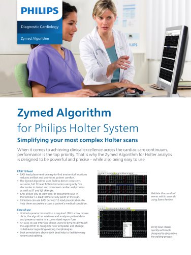 Zymed Algorithm Brochure
2 Pages
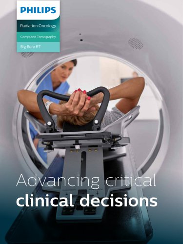 Big Bore RT Product Brochure
9 Pages
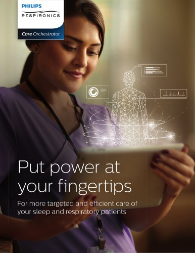 Care Orchestrator Brochure
8 Pages
Archived catalogs
 The time has come
24 Pages
 Philips InnerCool STx+
2 Pages
 Juno DRF Product Overview
4 Pages
 MDE Brochure
20 Pages
 MDE 3D-RX Brochure
6 Pages
 EasyDiagnost Eleva DRF
17 Pages
 Access Dual CT
8 Pages
 Philips ProGrade Product
4 Pages
 MobileDiagnost Opta
4 Pages
 Vereos PET/CT
8 Pages
 V60
V604 Pages
 HRC Mask Catalog - US
32 Pages
 MicroDose SI Product
16 Pages
 IntelliVue to Go
8 Pages
 Ingenuity TF PET/CT
20 Pages
 BrightView X and XCT
6 Pages
 Patients first performance
6 Pages
 PageWriter TC70
2 Pages
 HeartStart MRx
12 Pages
 Freedom of view
3 Pages
 Patients fi rst performance
6 Pages
 Critical Values
2 Pages
 fat-free imaging performance
2 Pages
 Azurion accessories
20 Pages
 Pinnacle3
6 Pages
 BV Pulsera
7 Pages
 Cardiology supplies guide
4 Pages
 Diamond Select
2 Pages
 Philips CardioMD IV
12 Pages
 ST80i stress-testing system
8 Pages
 Philips TPI
6 Pages
 PerforMax
2 Pages
 MobileDiagnost wDR
2 Pages
 Vereos Digital PET/CT brochure
21 Pages
 Compatibility table
4 Pages
 EPIQ 7
20 Pages
 dStream HeadNeckSpine coil
2 Pages
 Pinnacle³ Dynamic Planning
8 Pages
 Sparq - DS
4 Pages
 Trilogy 202
4 Pages
 DFM100
6 Pages
 DreamMapper
2 Pages
 MobileDiagnost wDR_2010
4 Pages


- Analysis software
- Philips Healthcare ultrasound system
- Trolley-mounted laser
- Philips Healthcare B/W ultrasound system
- Philips Healthcare color doppler ultrasound system
- Portable ultrasound system
- Philips Healthcare multipurpose ultrasound imaging system
- Visualization software
- Philips Healthcare medical imaging software
- Tablet computer software
- Tablet PC software
- Digital radiography system
- Reporting software
- Convex-array ultrasound system
- Linear-array ultrasound system
- Digital electrocardiograph
- EKG
- Multipurpose radiography system
- X-ray system
- Multi-parameter monitor