RAYSCAN-m-plus

RAYSCAN-m-plus
- 3D CBCT Applications: Utilized in otology, cochlear implants, neurotology, temporal bone analysis, rhinology, sinus surgery, and pediatric otorhinolaryngology.
- 2D Digital Radiography: Offers imaging for chest exams, laryngology, and skull and neck assessments with high-definition quality.
- High-resolution imaging allows precise diagnosis of small anatomical structures, such as the cochlea and auditory ossicles.
- Integration with ENT navigation systems enhances diagnostic and treatment planning.
- 3D printing capabilities for customized treatment solutions, such as cochlear implants and sleep apnea appliances.
- Reduced radiation dose compared to conventional CT scans through modulated cone beam width and pulsed X-ray technology.
- Visible light guide for precise area selection, minimizing unnecessary exposure.
- Motorized positioning and intuitive user interface for ease of operation.
- Supports various field-of-view (FOV) sizes for both 3D and 2D applications.
- Equipped with advanced detectors (CMOS, CdTe, a-Si TFT) for high-resolution imaging.
- Adjustable tube current and voltage settings to optimize image quality.
Catalog excerpts

3D Cone beam CT & Digital Radiography Dedicated to Otorhinolaryngology
Open the catalog to page 1
Multi-functional imaging solution RAYSCAN m is an unique 2-in-1 imaging solution, combining Cone Beam CT and Digital Radiography, designed for ENT specialists.
Open the catalog to page 2
3D CBCT applications - Otology and cochlear Implant - Neurotology and temporal bone - Rhinology and sinus surgery - Pediatric otorhinolaryngology 2D Digital radiography - Chest exam : PA / AP / Lateral - Laryngology - Skull: PA / AP / Lateral / Waters - Neck The state-of-the-art CBCT technology provides more accurate 3D images and 2D digital radiography options lead you to the best possible outcomes
Open the catalog to page 3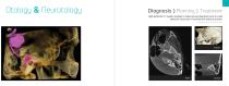
Otology & Neurotology Diagnosis Planning High definition CT quality enables to make precise diagnosis even on small anatomic structures of cochlea and auditory ossicles. Images are courtesy of SOREE Ear Clinic
Open the catalog to page 4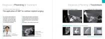
Diagnosis Planning Diagnosis Planning Diagnosis before implant surgery Case study of cochlear implant planning Follow-up after implant surgery The application of CBCT to cochlear implant surgery “An accurate measurement of the length of the cochlea is a selection of the optimal type of implant, which is essential for preserving residual hearing as maximally as possible.” “Using a high resolution cone beam CT, a line passing from the round window and the spiral center of the cochlea to its lateral wall can be correctly drawn. Thus, the length of the cochlea is measured.” Ray Digital solution I...
Open the catalog to page 5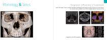
Rhinology & Sinus Diagnosis Planning Clear 3D images of sinus visualize detailed morphological information among air, bones and soft tissues. You can see more complete view of the anatomy which is not seen on 2D. Integration with ENT navigation
Open the catalog to page 6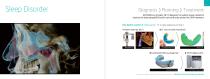
Sleep Disorder Diagnosis Planning RAYSCAN m provides 3D CT diagnosis for patient airway related to obstructive sleep apnea(OSA) which can be directly printed for OSA treatment. Ray Digital solution II : Sleep apnea CT to sleep appliance printing 1 Patient exam by 3D CT 2 CT scan of tooth information Airway diagnosis OSA Digital Design In progress of regulatory approval. Will be available in market soon. Opened to discuss business partnership
Open the catalog to page 7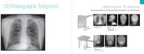
Medical grade 2D diagnosis Medical grade detectors provide high resolution images for each clinical practice. Neck : Lateral Skull : Waters - Foreign body aspiration - Lung condition - Epiglottitis, esophagus, trachea - Sphenoid, frontal, ethmoid adenoids, tonsils, cervical vertebrae - Maxillary sinus - Maxillary sinus Skull : Lateral Skull : Waters - Epiglottitis, esophagus, trachea - Sphenoid, frontal, ethmoid adenoids, tonsils - Maxillary sinus - Maxillary sinus Direct conversion
Open the catalog to page 8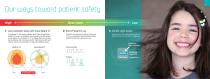
Our ways toward patient safety High Dose Level 1 Less radiation dose with Cone Beam CT Cone Beam CT has lower radiation dose than conventional medical CT exam, according to many known scientific papers. A key ability of cone beam CT is to change the field-of-view by modulating the cone beam width. Tight beam-width and shorter scans also contribute to reducing radiation doses. 2 Short Pulsed X-ray Pulsed X-ray operates to admit short pulse of X-ray into patient that relatively reduce radiation dose than continuous one. Low 3 Visible Light Guide Simply move the visible guiding light to select the...
Open the catalog to page 9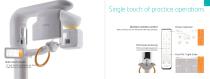
Single touch of practice operations Wireless remote control Protocol selection High sensitivity and non-directional make easy operation Motorized positioning Motorized height adjustment to set correct patient position Free FOV / Light Guide Wide touch screen - 10” wide monitor and intuitive user interface - Image preview to verify your exam
Open the catalog to page 10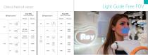
Clinical field-of-views Light Guide Free FOV 3D Applications
Open the catalog to page 11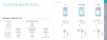
RAYSCAN m+ (Model: RCT710) Patient positioning Focal spot Tube current Tube voltage Specifications are subject to change without prior notice. Scan Ceph Detector type FOV / Image size Free FOV support Voxel / Pixel size Exposure time Standing (wheelchair accessible) 0.5mm 4~17mA 60~90kVp DR (Option) a-Si TFT Max. 42x42cm Yes 127pm Max. 3sec (0.2~0.8) Scan Ceph (Option) CdTe detector Max. 26x24cm Yes 100pm 4.9~9.9sec Operation Space Top View Front View
Open the catalog to page 12
Ray Co., Ltd. id 33Z-7, Samsungl-ro, Hwaseong-si, Gyeonggi-do, 18380, Korea Phone +82.31.605.1000 Email [email protected] Web www.raymedical.com Design and specifications are subject to change without notice. RBS-M02(rev.0)
Open the catalog to page 13All Ray catalogs and technical brochures
 ALL NEW RAYSCAN Brochure
11 Pages
 RIOScan
5 Pages
 RAYSCAN α+ Brochure
11 Pages
 RIOSensor Brochure
2 Pages
 RAYSCAN α Brochure
9 Pages
- Dental material
- Dental restoration material
- Resin dental material
- Tablet computer software
- Tablet PC software
- Flat panel detector
- Modeling dental material
- Dental software
- Design software
- 3D printer
- Dental radiography system
- Impression tray dental material
- 3D printing dental material
- Digital dental radiography system
- 3D scanner
- Temporary dental material
- Transparent dental material
- 3D dental scanner
- Dental 3D printer
- Desktop 3D printer