


inVia microscope
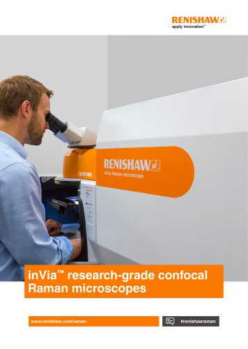
inVia microscope
The inVia™ confocal Raman microscopes by Renishaw are highly regarded for their performance and reliability, making them the top-selling Raman microscopes globally. Developed over two decades, these microscopes are durable and customizable for future needs, available in three models: inVia Basis, inVia Reflex, and inVia Qontor®.
- High Performance: Provides excellent data quality with high signal throughput and spectral resolution.
- Sensitivity: Detects weak Raman scatterers and analyzes thin films and monolayers.
- Flexibility: Configurable and upgradable for diverse sample analyses.
- Automation: Automates laser wavelengths, filters, and gratings, maintaining focus and alignment.
- Imaging Technologies: Offers comprehensive mapping and imaging techniques for detailed Raman images.
- LiveTrack™ Technology: Real-time focus tracking for uneven surfaces.
- Optical Efficiency: High efficiency with a stigmatic on-axis spectrometer.
- Spectral Resolution: Configurable for resolving narrow spectral features.
- Laser Options: Supports multiple lasers from near-infrared to deep ultraviolet.
- Detectors: Equipped with ultra-low noise, high sensitivity CCD cameras, accommodating up to four detectors.
- Material Analysis: Analyzes electronic and vibrational structures, measures material stresses.
- Sample Flexibility: Supports diverse configurations and accessories.
- Integration with Other Techniques: Combines with AFM, SEM, CLSM for comprehensive analysis.
Renishaw offers lifetime support and service, with remote diagnostics and on-site servicing. The inVia Raman microscope is designed for longevity and reliability, making it a valuable investment for research institutions.
The document highlights the inVia confocal Raman microscope's capabilities, including LiveTrack focus-tracking technology for real-time focus maintenance on rough surfaces.
Combines precise vertical motion control with optical focus-monitoring, allowing efficient Raman data collection from irregular surfaces and enabling 3D visualization of chemical and topographical data.
Supports advanced imaging techniques like StreamLine, StreamHR, and Rapide imaging, suitable for high spatial resolution and rapid data collection.
Renishaw’s WiRE software aids in Raman spectra acquisition and analysis, offering features like automatic background removal and noise reduction.
The inVia system is available in three models, each offering different levels of automation and features, with the Qontor model including full automation and LiveTrack technology.
Renishaw provides comprehensive training and support, including access to a global network of Raman application scientists and online training modules.
Details include spatial resolution, detector size, and operating temperature. The lateral spatial resolution is 0.25 µm, and the axial resolution is less than 1 µm. The standard detector size is 1024 x 256 pixels, operating at -70°C.
Controlled by a Windows® PC, with a supply voltage of 110 V AC to 240 V AC and a frequency of 50 Hz or 60 Hz. Typical power consumption is 150 W.
Performance varies with configuration. Contact Renishaw for detailed specifications.
Renishaw has been developing Raman spectroscopy products since 1992, ensuring reliable products through rigorous testing.
Details laser safety classifications according to IEC/EN 60825-1:2014, including Class 1, Class 3B, and Class 4 laser products.
Provides contact details for Renishaw, including phone number and email address.
Catalog excerpts
inVia™ research-grade confocal Raman microscopes www.renishaw.com/raman
Open the catalog to page 1
The world’s best selling high-performance Raman microscope • Designed, developed and refined over more than two decades to make it the most trusted Raman instrument on the market. • Superior, research-grade Raman microscope for your current and future needs. • Designed using Renishaw’s proven expertise in precision and innovative engineering. • Built to last: upgrade, reconfigure, or customise -the inVia Raman microscope is a sound investment. • Available in three models: inVia Basis; inVia Reflex and inVia Qontor®. • Many options and accessories are available to suit your analytical requirements...
Open the catalog to page 2
Why our users choose inVia Raman microscopes Renishaw is a global company with a worldwide network of scientists and engineers who are on-hand to provide you with expert product, technical and applications support Exceptional, reliable performance Quality, reliability and longevity The inVia system comprises a research-grade microscope coupled to a high-performance Raman spectrometer. It is simple to operate yet delivers outstanding performance—high signal throughput, combined with high spectral resolution and stability—giving reliable results, for even the most challenging measurements. The...
Open the catalog to page 3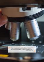
The performance of the system, together with the excellent support from Renishaw, made the decision [to buy an inVia] an easy one for us….. inVia microscopes are an efficient, easy-to-use, easy-to-share system. Boston University (USA) inVia™ confocal Raman microscop
Open the catalog to page 4
Key benefits High performance Repeatable results The inVia Raman microscope delivers outstanding performance, providing you with the best data in the shortest time. Rely on the inVia Raman microscope to produce results you can trust. With its outstanding performance you can be confident that it will deliver repeatable results time and time again – no matter how challenging the experiment. Sensitive See even the weakest Raman scatterers and get spectra from thin films and monolayers. Powerful Use the inVia Raman microscope for both Raman and photoluminescence measurements to obtain information...
Open the catalog to page 5
Key features High optical efficiency High spectral stability Fast and sensitive analysis Get consistent, reliable data Renishaw’s engineers have used their vast experience of precision and innovative design to make the inVia Raman microscope the most sensitive Raman instrument available. They use a stigmatic on-axis spectrometer which gives highest optical efficiency, excellent stray light rejection and exceptional sensitivity. With the inVia system you can study very weak Raman signals and rapidly analyse even minute traces of material. With its rigid, lightweight baseplate, and precision kinematic...
Open the catalog to page 6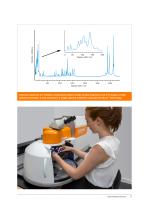
intensity / arbitrary A Raman spectrum of l-histidine, showing the lattice modes (inset), fingerprint and C-H ranges, at high spectral resolution. It was acquired in a single spectral collection using SynchroScan™ technology.
Open the catalog to page 7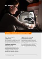
The inVia Raman microscope supports a wide range of environmental and sampling accessories. Here, a mapping experiment is being performed at high temperature. Highly sensitive detectors High performance microscope Cutting edge technology Leica for quality, efficiency and reliability The inVia Raman microscope's use Renishaw’s own ultra-low noise, ultra-high sensitivity CCD cameras so you get the best results in the shortest possible time. Should you wish to add more, the inVia system can be fitted with up to four detectors, such as electron multiplied (EM) detectors and InGaAs arrays. Leica Microsystems’...
Open the catalog to page 8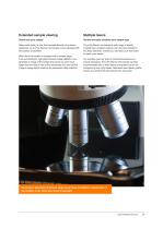
Extended sample viewing Multiple lasers Clearly see your sample Get the best data, whatever your sample type Many users prefer to view their samples directly using stereo eyepieces, so all inVia Raman microscopes come equipped with binoculoars as standard. The inVia Raman microscope’s wide range of directly coupled laser excitation options, from the near-infrared to the deep ultraviolet, ensures you can tailor your instrument to match your needs. When the inVia system is equipped with a sample stage, such as Renishaw’s high speed encoded stage (MS30), it can generate an image of the sample that...
Open the catalog to page 9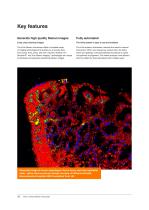
Crisp, clear chemical images The inVia Raman microscope offers a complete range of imaging technologies that enable you to acquire data from points, lines, areas, and even volumes. StreamLine™, StreamHR™ and True Raman Imaging™ technologies are unique to Renishaw and generate outstanding Raman images. The inVia system is easy to use and maintain The inVia system’s automation removes the need for manual intervention. When you change key components, like filters, lasers and gratings, it will automatically reconfigure its optics and optimise its alignment. This makes analysis more efficient, which...
Open the catalog to page 10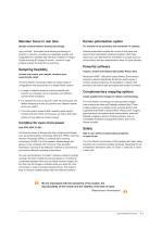
Maintain focus in real time Raman polarisation option Sample surface/interface tracking technology For analysis of the symmetry and orientation of samples Use LiveTrack™ automated focus-tracking technology to acquire, in real-time, accurate and repeatable spectra and topography from samples with extensive variations in height. Create stunning 3D images of uneven, curved or rough surfaces without the need for pre-scanning. Optional polarisation enables the control of both laser and spectrometer polarisation (polariser/analyser). With these options you can determine the orientation of crystals...
Open the catalog to page 11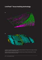
(a) Analysis of a coated flat-head screwdriver bit. The image reveals the distributions of TiN (green) and TiO2 (magenta). The imaged surface is 8.1 mm wide, 5.1 mm deep and 3.6 mm high. (b) Quartz-dominated rock (Tiger’s Eye). The Raman image shows quartz (cyan) and inorganic carbonates (yellow). The imaged surface is 47 mm wide, 26 mm deep, and 3.0 mm high. 12 inVia™ confocal Raman microscope
Open the catalog to page 12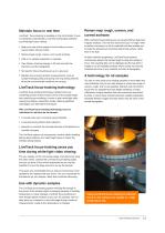
Maintain focus in real time LiveTrack focus-tracking is available on the inVia Qontor. Focus is maintained automatically in real time during data collection and white-light video viewing. ™ • Keep your view of the sample in focus while you move around under manual control. • Raman-image rough, uneven, and curved surfaces. • Little or no sample preparation is required. • View Raman chemical images in 3D and see both the chemistry and the topography. • No need for time-consuming set up or a pre-scan. • Maintain focus during dynamic measurements, such as sample heating/cooling and during very long...
Open the catalog to page 13All Renishaw catalogs and technical brochures
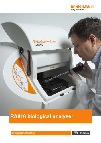 RA816 Biological Analyser
10 Pages
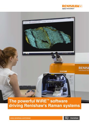 WiRE™ software
14 Pages
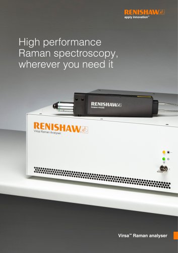 Virsa™ Raman analyser
12 Pages
Archived catalogs
 WiRE™ software
8 Pages
 inVia™ confocal Raman microscope
28 Pages
 3D modelling and printing
4 Pages
 Mission to Nepal
4 Pages
 Renishaw CMM fixtures
36 Pages
 LaserImplant™
1 Page
 Brochure : Delivering precision
16 Pages
 Brochure neuromate
16 Pages
 LaserAbutments™
4 Pages
 M6 CMM clamping kit
2 Pages
 DS10 system specifications
2 Pages
 Neuroguide leaflet
2 Pages
 neuromate® stereotactic robot
17 Pages


- Analysis software
- Compound microscope
- Laboratory microscope
- Desktop microscope
- Control software
- Laboratory testing system
- Windows software
- Laboratory software
- Planning software
- Automated software
- Acquisition software
- Surgery software
- Analyser for the pharmaceutical industry
- Benchtop spectrometer
- Research microscope
- Binocular microscope
- High-resolution microscope
- Data processing software