Measuring mitochondrial membrane potential with JC-1 using the Cellometer Vision image cytometer.
1 /4Pages

Measuring mitochondrial membrane potential with JC-1 using the Cellometer Vision image cytometer.
1 /4Pages
Catalog excerpts
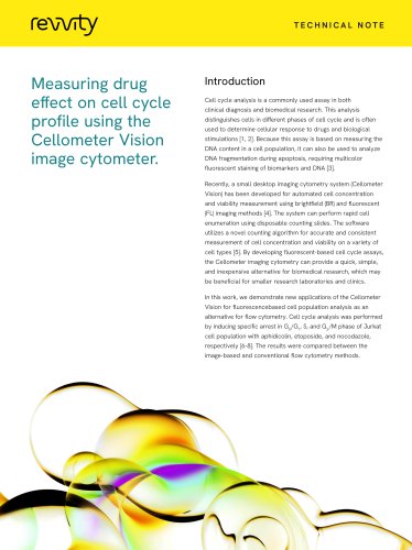
TECHNICAL NOTE Measuring mitochondrial membrane potential with JC-1 using the Cellometer Vision image cytometer. Introduction Alterations in apoptosis are relevant to a large number of disease states, including cancer, heart disease, neurological disorders, infectious disease, and more [1]. One of the earliest steps in the apoptosis pathway, the loss of mitochondrial membrane potential, is often an indication of disorder and is therefore a useful target for those investigating the mechanism of disease in vitro. Mitochondrial membrane potential can be easily and efficiently monitored using a number of commonly available fluorescent dyes. The Cellometer® Vision, a small desktop image cytometry system has been developed for automated brightfield (BR) and fluorescent (FL) imaging methods [2]. The system can perform rapid cell enumeration and other fluorescent measurements using disposable counting slides. The software utilizes a novel counting algorithm for accurate and consistent measurements on a variety of cell types [3]. By developing fluorescent-based assays to assess mitochondrial membrane potential, the Cellometer imaging cytometry can provide a quick, simple, and inexpensive alternative for biomedical research, which may be beneficial for smaller research laboratories and clinics. In this work, we demonstrate a mitochondrial membrane potential assay using Cellometer imaging cytometry and the JC-1 dye as an alternative to flow cytometry. The data obtained by Cellometer were compared to those from conventional flow cytometry methods.
Open the catalog to page 1
Measuring mitochondrial membrane potential with JC-1 using the Cellometer Vision image cytometer. Materials and methods Cellometer Vision and disposable counting chamber Cell preparation for membrane potential analysis The Cellometer Vision utilizes one brightfield and two The MitoProbe™ JC-1 assay kit was purchased for JC-1 fluorescent channels to perform image-based cytometric mitochondrial membrane potential analysis. The assay analysis. Brightfield imaging used a broadband white measures the depolarization of mitochondrial membrane light-emitting diode (LED) and fluorescence imaging used...
Open the catalog to page 2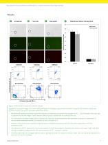
Measuring mitochondrial membrane potential with JC-1 using the Cellometer Vision image cytometer. Red/Green Ration Comparison • Cellometer S Flow Cytometer Flow Cytometer Figure 1: Mitochondrial membrane potential analysis. Brightfield, fluorescent images, and scatter plots generated by Cellometer (top) and flow cytometer (bottom) of (A) unstained Jurkat cells, (B) control Jurkat cells, and (C) cells with induced with CCCP. • Mitochondrial membrane potential experiment required two fluorescence detection wavelengths for JC-1. The Cellometer Vision was able to generate fluorescent images in both...
Open the catalog to page 3
Measuring mitochondrial membrane potential with JC-1 using the Cellometer Vision image cytometer. • The ability to rapidly and cost-effectively perform 1. Favaloro, B., et al., Role of apoptosis in disease. imaged-based mitochondrial membrane potential assays may improve research efficiency, especially where a flow or laser scanning cytometer is not available or in Aging (Albany NY), 2012. 4(5): p. 330-49. 2. Chan, L.L., et al., Direct concentration and viability measurement of yeast in corn mash using a novel situations where a rapid analysis of data is critical. imaging cytometry method. J...
Open the catalog to page 4All Revvity catalogs and technical brochures
 Model 307 Sample Oxidizer
3 Pages


Archived catalogs
 chemagic 360 instrument
4 Pages
 chemagic Prepito® instrument
4 Pages


- Detection kit
- Solvent reagent
- Blood detection kit
- Molecular biology reagent
- Serum detection kit
- Immunoassay detection kit
- Plasma detection kit
- Research reagent
- Laboratory reagent
- Diagnostic reagent
- Infectious disease detection kit
- Molecular detection kit
- Optical test kit
- Clinical detection kit
- Sample preparation system
- Tablet computer software
- Tablet PC software
- Automatic sample processor
- Microtiter plate
- Fluorescence detection kit