- Catalogs
- Richard Wolf
- VERTEBRIS cervical Brochure
- Products
- Catalogs
- News & Trends
- Exhibitions
VERTEBRIS cervical Brochure

VERTEBRIS cervical Brochure
This document explores the development and techniques of full-endoscopic decompression of the cervical spine, focusing on both posterior and anterior approaches. It traces the evolution of surgical procedures from the 1940s and 1950s, discussing the benefits and challenges of anterior decompression and fusion, as well as posterior foraminectomy.
Storage and Access Determination: The patient is positioned prone with the cervical spine in a fixed lordotic position. Access is determined using anatomical landmarks and image intensifier control.
Implementation and Operating Procedure: A stab incision is made, and a dilator is inserted to contact the bone. The endoscope is used through a working sleeve, and bone resection is performed to expose neural structures and remove spinal disk herniations.
Storage and Access Determination: The patient is placed supine with the cervical spine slightly reclined. Access is made contralaterally to the pathology, using image intensifier control.
Implementation and Operating Procedure: A dilator is inserted into the intervertebral space, and the endoscope is used through a surgical sleeve. Bone resection is often necessary to access the spinal canal and remove herniations.
The document details the instruments used for full-endoscopic procedures, including endoscopes, dilators, and working sleeves. It also mentions optional instrumentation and systems like the Radioblator RF and PowerDrive ART1.
The document emphasizes the importance of avoiding manipulation of the spinal cord and highlights the potential risks and complications associated with cervical spine surgeries. It stresses the need for skilled surgeons and the availability of equipment for open surgery if necessary.
- Working Instruments: Includes elevators, dissectors, probes, reamers, punches, and rongeurs with specific outer diameters (OD) and working lengths (WL).
- Auxiliary Instruments: Features atraumatic elevators, sharply abrading reamers, and bone punches.
- Color Coding: Instruments are color-coded for easy identification based on diameter.
- TipControl RF System: Comprises bipolar handles, sheath tubes, and connection cables for RF devices. It is designed for full-endoscopic spine surgery.
- Radioblator RF 4 MHz: A multidisciplinary system offering monopolar and bipolar cutting and coagulation modes with user presets.
- VERTEBRIS Cervical Instrumentation: Includes both anterior and posterior techniques with specific endoscopes, telescopes, and fiber light cables.
- Access Systems: Designed for instruments with maximum OD specifications, including dilators, working sleeves, and guide cannulas.
- PowerDrive ART1: A universal motor system with features like automatic handle and tool recognition, and user-specific parameter storage.
- Motor Handles: Power Stick M5 with rotary speed options and operation via foot switch or touchpad.
- FLUID CONTROL Arthro-Spine: An irrigation and suction pump system with automatic tube recognition, designed for arthroscopy and full-endoscopic spine surgery.
- Accessories: Includes reusable and single-use tube sets, vacuum tubes, and suction containers.
- TipControl RF Instruments: Includes sheath tubes and electrodes for radiofrequency surgical systems.
- Single-use Access Instruments: Spinal cannula sets and drain tubes for surgical procedures.
Catalog excerpts

VERTEBRIS cervical Full-endoscopic Decompression of the cervical spine posterior and anterior techniques
Open the catalog to page 1
RICHRRD nrin VERTEBRIS cervicalFull-endoscopic Spine Instrumentation spirit of excellence Contents VERTEBRIS cervical The full-endoscopic posterior technique ■ Storage i Determination of the access ■ Implementation of the access i Operating procedure The full-endoscopic anterior technique ■ Storage i Determination of the access ■ Implementation of the access i Operating procedure VERTEBRIS cervical Instrumentation ■ VERTEBRIS cervical posterior 14 i VERTEBRIS cervical anterior 16 ■ Radioblator RF 4 MHz - Multidisciplinary Radiofrequency Surgical System 20 ■ PowerDrive ART1 - Universal...
Open the catalog to page 3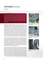
VERTEBRIS cervical Foreword In the area of the cervical spine, radicular symptoms due to degenerative causes, in other words pain in the arms, are typically caused by mediolateral to lateral spinal disk herniations or stenoses of the intervertebral foramen. At the beginning of the 1940s, the clinical symptoms of this nature with a topographical reference to changes in the cervical disks were classified for the first time. Although good results are frequently obtained using conservative methods, surgical intervention may become necessary in the presence of pain or neurological deficits. The development...
Open the catalog to page 4
the spinal column - and different surgical instruments provide technical conditions akin to conventional microscopically assisted surgical inventions with the simultaneous advantages of the full-endoscopic approach with 25° telescopes with a continuous flow of fluid.1 The main indications for cervical full-endoscopic operations are "soft" spinal disk herniations with radicular symptoms, in other words pain in the arms. Since the cervical spinal cord cannot be manipulated medially, the posterior approach is used for herniations where the main section is localized laterally to the lateral edge...
Open the catalog to page 5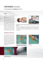
VERTEBRIS cervical The full-endoscopic posterior technique Storage The operation is performed with the patient in the prone position lying on a hip and thorax roll. The head and the cervical spine must be resting with correct lordotic adjustment in a fixed position in keeping with a posterior intervention on the cervical spine. X-ray monitoring should be permitted during the operation in two planes. General fixation in the Mayfield Clip or a similar holder offers excellent prerequisites and always provides the circumstances for an open Prone position, fixation of the head in the Mayfield Clip,...
Open the catalog to page 6
Implementation of the access After determining the entry point in the skin and carrying out a stab incision, the dilator is inserted until contact is made with bone on the zygoapophyseal joint under lateral image intensifier control. The working sleeve with oblique opening is pushed through the dilator in a medial direction and the dilator is removed. Insertion of the dilator in the zygoapophyseal joint The operating sleeve is inserted through the dilator Operating procedure The endoscope is inserted through the working sleeve. The operation is carried out in vision using different instrument...
Open the catalog to page 7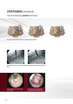
Bony parts of the joint and the lamina are resected to open the foramen The image intensifier can help with orientation during cutting or when working in the spinal canal Reamed foramen with view of the liga- View in the lateral spinal canal with mentum flavum cervical spinal cord and spinal nerve
Open the catalog to page 8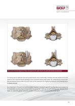
After removal of the lateral ligamentum flavum and exposure of the neural structures, the spinal disk herniation can be removed. The locking caps for telescope and working sleeve should only be used briefly if bleeding obscures visibility since when operations last a long time and the drainage of fluid is prevented without being noticed, the consequences of volume overload and elevated pressure within the spinal canal and the associated and neighboring structures cannot theoretically be completely excluded. Any manipulation of the spinal cord must be avoided. Experience indicates that generally...
Open the catalog to page 9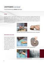
VERTEBRIS cervical The full-endoscopic anterior technique Storage The patient is placed in a supine position. The head and cervical spine must be placed in a slightly reclining position and fixed in keeping with an anterior approach to the cervical spine. X-ray monitoring should be permitted in two planes during the procedure. General fixation in the Mayfield Clip or a similar holder offers excellent prerequisites and always provides the circumstances for an open intervention if an emergency occurs. Particularly in the case of the lower cervical spine, it may be necessary to tape the shoulders...
Open the catalog to page 10
Implementation of the access After determining the entry point in the skin and carrying out a stab incision in the skin, the first thin dilator is inserted in the intervertebral space under lateral image intensifier control. It is important to make an anterior puncture in the spinal disk and not to miss laterally. This not only precludes further operations but can also lead to injury of the vertebral artery, spinal nerve or esophagus. Alternatively, the spinal disk can be punctured using a spinal cannula and a guide wire is inserted through this. The first dilator can then be pushed through this....
Open the catalog to page 11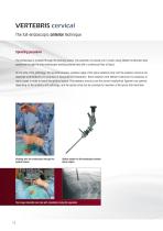
VERTEBRIS cervical The full-endoscopic anterior technique Operating procedure The endoscope is inserted through the working sleeve. The operation is carried out in vision using different instrument sets positioned through the intra-endoscopic working channel and with a continuous flow of liquid. On the side of the pathology, the uncinate process, posterior edge of the spine vertebral body and the posterior annulus are dissected contralaterally for purposes of topographical orientation. Bone resection with different instrument is necessary in many cases in order to reach the epidural space. The...
Open the catalog to page 12
Bony resection is frequently necessary to reach the spinal canal Depending on the findings, the dorsal longitudinal ligament should be opened The locking caps for the telescope and the working sleeve should only be used at short notice for hemorrhage that obscures vision because if operations extend over a long period and if the drainage of the irrigation fluid is inadvertently obstructed the consequences of volume strain and pressure increase cannot be entirely excluded within the spinal canal and the associated and adjacent structures. Any manipulation of the cervical spinal cord must be avoided....
Open the catalog to page 13All Richard Wolf catalogs and technical brochures
 FLUID CONTROL 2225
12 Pages
 Catalogue Urology
466 Pages
 Catalogue Orthopedics
218 Pages
 Catalogue OR Integration
36 Pages
 Catalogue Gynecology
248 Pages
 Catalogue General surgery
222 Pages
 Catalogue Traitement
58 Pages
 Sterilization Basket System
8 Pages
 ERAGON modular
28 Pages
 ENT catalogue
426 Pages
 LEVD
LEVD8 Pages
 COXARTIS Brochure
12 Pages
 BioactIF OSTEOTRANS Brochure
4 Pages
 ERAGONbipolar Folleto
4 Pages
 MegaPulse 70+ Flyer
2 Pages
 RIWO-System-Tray Brochure
4 Pages
 ERAGON
8 Pages
 PANOVIEW ULTRA
4 Pages
 Fiber light cables
4 Pages
 ENDOCAM Performance HD
3 Pages
 Endocam Logic HD
12 Pages
 ENDOLIGHT LED
6 Pages
 VERTEBRIS denervation
4 Pages
 FLUID CONTROL Arthro
4 Pages
 VERTEBRIS lumbar-thoracic
56 Pages
 ENDOCAM Flex HD
12 Pages
 Disposables by Richard Wolf
2 Pages
 core nova
8 Pages
 ZeroWire Brochure
2 Pages
 ENDOCAM Epic 3DHD Brochure
8 Pages
 core.media Brochure
2 Pages
 core nova Brochure
8 Pages
 core nova Image Brochure
2 Pages
 Sterisafe® DURO A3
4 Pages
 Steri Baskets Prospekt
8 Pages
 Eragon Modluar Mini
8 Pages
![Catalogue [Translate to English:] Disposables Urology](https://img.medicalexpo.com/pdf/repository_me/78958/catalogue-translate-to-english-disposables-urology-196889_1mg.jpg)


Archived catalogs
 B 778 Lap Bergebeutel I13 GB
2 Pages
 B 785 Titanium Clip GB VII13
2 Pages
 B 784 Eragon axial GB II13
4 Pages
 B 920 TEM D GB VI10
18 Pages
 B 775 LEVD XI12 GB
6 Pages
 B 760 ERAGON bipolar GB VI10
4 Pages
 B 773 ERAGON modular GB VII11
12 Pages
 B 772 Minilap GB IV11
4 Pages
 B 770 ERAGON compact GB VI11
12 Pages
 B 774 Key Port I13 GB
12 Pages
 Reprocessing baskets
2 Pages
 Sterisafe DURO A3 Brochure
4 Pages
 VictOR HD Brochure
6 Pages
 RECTO PUMP Brochure
2 Pages
 arthroLution Folleto
4 Pages
 TipControl Brochure
4 Pages
 PowerDrive ART1 Brochure
8 Pages
 T-Lock OSTEOTRANS
6 Pages
 COMBIDRIVE EN Brochure
20 Pages
 Ultra Thin Uretero-Renoscope
2 Pages
 FLUID CONTROL LAP Brochure
2 Pages


- Surgery forceps
- Grasping forceps
- Trolley-mounted laser
- Surgery electrode
- Human scissors
- Tabletop laser
- Medical endoscope
- Surgical scissors
- Visualization software
- Orthopedic surgery instrument kit
- Control software
- Endoscopy forceps
- Electrosurgical forceps
- Needle holder
- Surgical needle holder
- Sealing forceps
- CMOS camera
- Camera with USB port
- Tissue grasping forceps
- Medical imaging display