- Catalogs
- RIGEL Medical
- Rigel Guide Defibrillation and Transcutaneous Pacing 2021
- Products
- Catalogs
- News & Trends
- Exhibitions
Rigel Guide Defibrillation and Transcutaneous Pacing 2021
1 /32Pages

Rigel Guide Defibrillation and Transcutaneous Pacing 2021
1 /32Pages
Catalog excerpts

An Introduction to Defibrillation and Transcutaneous Pacing Passionate about patient safety.
Open the catalog to page 1
AN INTRODUCTION TO DEFIBRILLATION AND TRANSCUTANEOUS PACING Defibrillator Devices Analysing Defibrillator Energy Products in the Rigel Medical Range Accessories and Services from Rigel Medical Rigel Medical: Keeping medical industries safe World-leading biomedical test and measurement equipment. At Rigel Medical, we’ve been a pioneer of biomedical test and measurement instruments for more than four decades. Since launching the world’s first IEC 601 electrical safety analyzer in the 1970s, our user focused developments continue to place us at the forefront of the industry. Today, our UniPulse...
Open the catalog to page 2
Defibrillator testing made easy... ...with the UniPulse 400 defibrillator analyzer. Passionate about patient safety. Designed to comprehensively test all defibrillators quickly and accurately, the UniPulse 400’s large colour screen displays test results and waveforms in amazing detail, while dedicated fast keys and easy to use operating system enables engineers to select the right test function in seconds. Features include: • Light handheld, battery operation • Reliable and non-inductive test load of 50Ω • Pacer functionality • Compatible with monophasic, biphasic and pulsed waveforms • Patient...
Open the catalog to page 3
AN INTRODUCTION TO DEFIBRILLATION AND TRANSCUTANEOUS PACING This booklet is written as a guideline for people involved in testing medical defibrillator devices. All reasonable care has been taken to ensure that the information, reference figures and data are accurate and have been taken from the latest versions of various standards, guidance notes and recognised best practices to establish the recommended testing requirements. Rigel Medical, their agents and distributors, accept no responsibility for any error or omissions within this booklet or for any misinterpretations by the user. A cardiac...
Open the catalog to page 4
Passionate about patient safety. Figure 1 – Advanced life support external defibrillator Internal defibrillators – implantable cardioverter defibrillators (ICD) - are similar to pacemakers, monitoring the heart’s electrical activity to deliver a shock if needed. An external defibrillator can be operated to deliver a shock, either monophasic or biphasic, to the patient by a physician, a paramedic, or an untrained bystander. Automated external defibrillators (AEDs) - often referred to as public access defibrillators (PADs) - are easy to use and are becoming increasingly common, typically found...
Open the catalog to page 5
AN INTRODUCTION TO DEFIBRILLATION AND TRANSCUTANEOUS PACING History With the advances in medicine at the turn of the 20th century, heart disease was a serious health concern due to people living longer, and became the leading cause of death. People had usually died from infectious diseases before reaching an age where heart problems could threaten their health. Around this period, Swiss researchers JeanLouis Prévost and Frédéric Batelli brought about the paradox that electric current could lead to fibrillation of the cardiac chambers, and more importantly defibrillation, when experimenting on...
Open the catalog to page 6
Passionate about patient safety. Figure 2: Kouwenhoven’s open chest defibrillator [10]
Open the catalog to page 7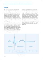
AN INTRODUCTION TO DEFIBRILLATION AND TRANSCUTANEOUS PACING Heart The heart is located in the thoracic cavity. It pumps blood, creates blood pressure, and circulates oxygen, nutrients and other substances we absorb around our body. The heart has four chambers: the right and left atria, and the right and left ventricles. The chambers are made up of cardiac muscle known as myocardium. The electrical activity of the myocardium regulates the mechanical sequence of events known as the cardiac cycle (Figure 3). In its simplest form, the cardiac cycle is the simultaneous contraction (systole) of the...
Open the catalog to page 8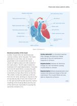
Passionate about patient safety. Electrical activity of the heart Cardiac muscle cells can spontaneously contract without nerve impulses. The heart generates its own beat, and its natural pacemaker is the sinoatrial (SA) node – a cluster of highly conductive cells located in the right atrium. The SA node initiates the heartbeat with rapid depolarisation. Impulses from the SA node follow a conductive path to the AV node and atrial myocardium; this transmission brings about atrial contraction. The cardiac conductive pathway continues from the AV node, sending an impulse to the AV bundle (bundle...
Open the catalog to page 9
AN INTRODUCTION TO DEFIBRILLATION AND TRANSCUTANEOUS PACING Figure 5: ECG electrical activity Electrocardiogram The series of electrical events generated by the myocardium can be depicted using an electrocardiogram (ECG) monitor when electrodes are attached to the skin across the chest, arms and legs. A typical ECG (Figure 5) consists of: the P wave, representing SA node depolarisation of the atria; the QRS complex, representing depolarisation on the ventricles as the impulses distribute through the ventricular myocardium; and the T wave, representing the repolarisation of the ventricles. Atria...
Open the catalog to page 10
Passionate about patient safety. Figure 6: Refractory period Types of arrhythmia are: ∙ Atrial Fibrillation (AF) - rapid irregular contraction of the atria ∙ Atrial Flutter (AFL) - similar to AF but not irregular ∙ Bradycardia - slow resting heart rate <60bpm ∙ Ventricular Tachycardia (VT) - fast resting heart rate >100bpm ∙ Ventricular Fibrillation (VF) disorganised electrical signals cause the ventricles to rapidly quiver Fibrillation is very rapid, uncoordinated contractions that cause a sudden reduction in cardiac output (the amount of blood pumped by the ventricles per minute). The ventricles...
Open the catalog to page 11
AN INTRODUCTION TO DEFIBRILLATION AND TRANSCUTANEOUS PACING Defibrillation Defibrillation is the corrective measure to stop VF or, to simplify, convert arrhythmia to a NSR. A defibrillator can deliver a strong electric shock to the heart, simultaneously depolarising the cardiac cells so they contract. The cells then simultaneously repolarise themselves to a relaxed state. A normal heart beat will “restart” if the first part of the heart to recover is the SA node. The energy delivered to a patient must be adequate to affect the heart cells. In general, short duration shocks require larger currents,...
Open the catalog to page 12All RIGEL Medical catalogs and technical brochures
 Med-eBase
2 Pages
 SafeTest 60
3 Pages
 CITREX H3
1 Page
 SafeTest 99
3 Pages
 FlowAnalyser PRO
4 Pages
 CITREX H5
2 Pages
 Guide to Infusion Pump Testing
19 Pages
 Guide to Electrosurgery
19 Pages
 IEC 62353
29 Pages
 IEC 60601
18 Pages
 Global Brochure
17 Pages
 CITREX
6 Pages
 PRODUCT RANGE 2017
12 Pages
 Printers and Scanners
6 Pages
 Rigel 62353+
5 Pages

Archived catalogs
 UniPulse 400
2 Pages
 VenTest 800 Series
8 Pages
 Rigel 288+
5 Pages
 PULS-R
2 Pages
 PRODUCT RANGE 2018
12 Pages
 PatSim200
2 Pages
 Rigel Multi-Flo Datasheet
6 Pages
 RIGEL
RIGEL20 Pages
 SP-SIM SpO2 Simulator
5 Pages
 BP-SiM NIBP Simulator
5 Pages
 M e d - e B a s e v 2 . 0
3 Pages
 Rigel Medical Brochure - USA
20 Pages
 Rigel Medical Brochure UK
20 Pages
 Uni-Pulse
6 Pages


- Analysis software
- Control software
- Avalue laboratory tester
- Monitoring software
- Medical gas flow meter
- Shoulder bag
- Hospital flow meter
- Avalue medical device tester
- Avalue control tester
- Patient simulation unit
- Data management software
- Thermal printer
- Measurement software
- Medical device bag
- IBP cable
- Temperature testing system
- Portable simulation unit
- Recording software
- Monitoring simulator
- Medical device battery