- Catalogs
- RISystem AG
- MouseNail
- Company
- Products
- Catalogs
- News & Trends
- Exhibitions
MouseNail
1 /27Pages
MouseNail
1 /27Pages
Catalog excerpts

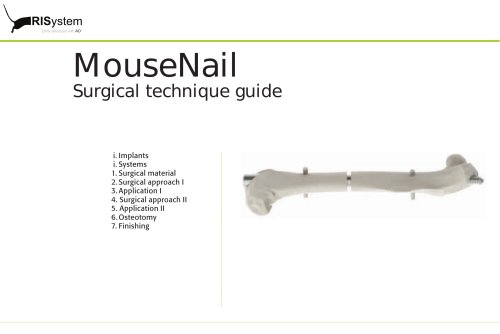
“jointly developed with AO” Surgical technique guide i. Implants i. Systems 1. Surgical material 2. Surgical approach I 3. Application I 4. Surgical approach II 5. Application II 6. Osteotomy 7. Finishin
Open the catalog to page 1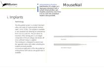
“jointly developed with AO” Intramedullary fixation Stabilization of a single cut / large gap osteotomy with a locked intramedullary nail. Possible osteotomy sizes from 0.25 to 2.00 mm. A fracture model is also possible. Technology The MouseNail system is a locked intramedullary nail made of medical grade stainless steel 1.4441 (316L). The implant is available in one standard size allowing for osteotomies from 0.25 to 2.00 mm. The pin driver for insertion of the locking pins is an integral component of the pin design and shears off once sufficient torque is applied. The locking pin is designed...
Open the catalog to page 2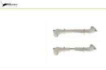
“jointly developed with AO” i. Systems The MouseNail system can cover single cut osteotomies up to large bone defects within the femur. MouseNail with 0.25 mm osteotomy MouseNail with 2.00 mm osteotom
Open the catalog to page 3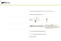
“jointly developed with AO” 1. Surgical material Implants: - 1x MouseNail - 2x MouseNail locking pin RIS.221.122 MouseNail wide interlocking Implant specific instruments: - 1x MouseNail aiming device - 1x Saw guide 0.25 mm RIS.221.201 MouseNail aiming device, wide interlocking Instruments: - 4x hand drills - 1x Accu Pen 3V RIS.390.130 Hand drill RIS.390.200 AccuPen 3V Consumables: - 1x 0.30 mm Drill bit - 1x 0.22 mm Gigly wire saw, 0.50 m - 1x 1.00 mm centering bit - 1x 22-G/27-G needle, length > 19 mm - 1x Vicryl suture 3-0 & Vicryl suture 5-0 - 1x Ethibond Vicryl suture 6-0
Open the catalog to page 4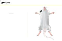
“jointly developed with AO” 2. Surgical approach I Positioning Mouse in dorsal position.
Open the catalog to page 5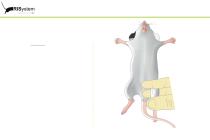
“jointly developed with AO” 2. Surgical approach I Positioning The knee is bent according to illustration.
Open the catalog to page 6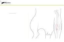
“jointly developed with AO” 2. Surgical approach I Dorsal approach Longitudinal incision along the medial side of the patella from the distal third of the thigh to the proximal third of the lower leg.
Open the catalog to page 7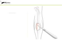
“jointly developed with AO” 2. Surgical approach I Dorsal approach Longitudinal incision along the medial side of the patellar tendon.
Open the catalog to page 8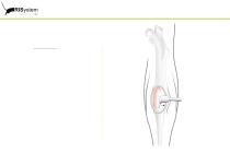
“jointly developed with AO” 2. Surgical approach I Dorsal approach Lateral dislocation of the patella to expose the knee joint.
Open the catalog to page 9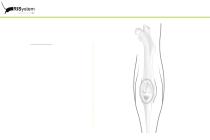
“jointly developed with AO” 2. Surgical approach I Dorsal approach Position of the intercondylary entry point of the femur.
Open the catalog to page 10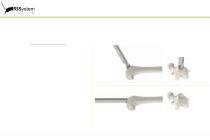
“jointly developed with AO” 3. Application I Opening of the epicondyle Use the 1.00 mm centring bit to drill a hole into the intercondylar notch. According the illustration start drilling with a 45 ° offset to the axis of the femur and continuously decrease the angle to 0° offset (parallel with the bone axis). Make sure not to exceed 2.00 mm in depth for the drill hole ! Verify the orientation of the longitudinal axis and keep the centering bit right between the two condyles of the medullary cavity and parallel to the bone axis.
Open the catalog to page 11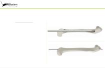
“jointly developed with AO” 3. Application I Proximal opening of femur Use a 22G needle (length > 19 mm) to ream the medullary cavity. Again make sure that the needle is central to the medullary cavity and parallel to the bone axis ! Open the femur proximally with a 27G needle (length > 19mm) to prepare the insertion of the MouseNail.
Open the catalog to page 12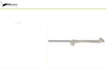
“jointly developed with AO” 3. Application I Insertion Insert the MouseNail under continous rotation until the distal end of the MouseNail is flush with the epycondyle. Make sure to apply smooth axial presure during insertion.
Open the catalog to page 13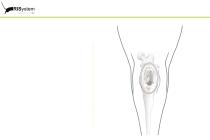
“jointly developed with AO” 3. Application I MouseNail in situ.
Open the catalog to page 14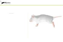
“jointly developed with AO” 4. Surgical approach II Positioning Mouse in lateral position.
Open the catalog to page 15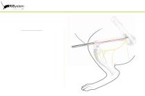
“jointly developed with AO” 4. Surgical approach II Anterolateral approach Longitudinal incision along the femur from the hip joint to the knee.
Open the catalog to page 16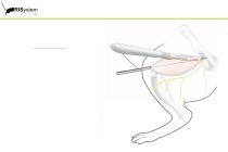
“jointly developed with AO” 4. Surgical approach II Anterolateral approach Longitudinal incision of the fascia lata.
Open the catalog to page 17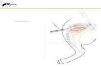
“jointly developed with AO” 4. Surgical approach II Anterolateral approach M. vastus lateralis and M. biceps femoris are split and M. tensor fasciae latae is lifted to expose the full length of the femur (preserving the sciatic nerve).
Open the catalog to page 18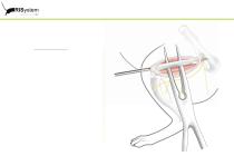
“jointly developed with AO” 4. Surgical approach II Anterolateral approach Circular preparation of the femur.
Open the catalog to page 19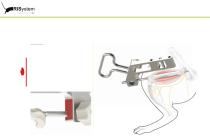
“jointly developed with AO” 5. Application II Assembly Mount the aiming device to the MouseNail. Pay special attention to the final position of the aiming device. If there is a gap between the MouseNail adapter flanch and the inner surface of the aiming device (marked in red) than the aiming device needs to be advanced more until the stop on the MouseNail and the inner surface get in contact.
Open the catalog to page 20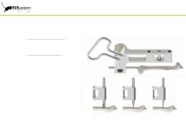
“jointly developed with AO” 5. Application II Orientation of the aiming device Position the aiming device anterolaterally to the femur. Interlocking of the MouseNail Starting with the more distal interlocking position use the 1.00 mm centering bit to prepare for drilling. Maintain the orientation of the aiming device and drill the first interlocking hole with the 0.3 mm bit. The countersinking as well as the drilling should be done by hand without the use of power tools ! Carefully apply the MouseNail locking pin via the aiming device and bring it into its final position by turning it into the...
Open the catalog to page 21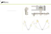
“jointly developed with AO” 6. Osteotomy Performing the osteotomy Attach the 0.25 mm saw guide on the lateral side of the aiming device and create a defined gap by using the Gigly saw (sufficient irrigation !) To avoid damage of the soft tissue cut the saw wire close to the bone on either side.
Open the catalog to page 22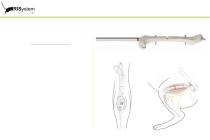
“jointly developed with AO” 6. Osteotomy Accomplishing the osteotomy Remove the aiming device from the MouseNail and disconnect the shaft of the MouseNail at the groove (marked red).
Open the catalog to page 23All RISystem AG catalogs and technical brochures
 MouseScrew
16 Pages
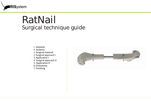 RatNail
25 Pages
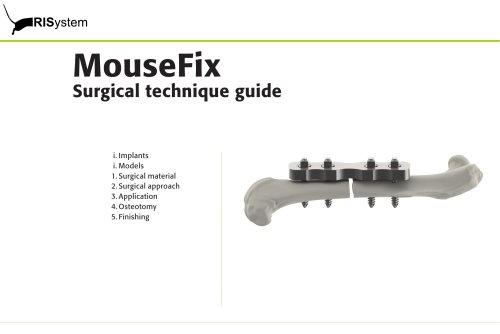 MouseFix
17 Pages
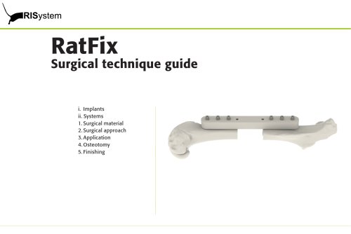 RatFix
15 Pages
- Veterinary instrument kit
- Compression veterinary orthopedic screw
- Veterinary bone plate
- Orthopedic surgery veterinary instrument kit
- Tibia veterinary bone plate
- Mice veterinary bone plate
- Rat veterinary bone plate
- Small animal veterinary instrument kit
- Veterinary intramedullary nail
- Femur veterinary intramedullary nail
- Femur veterinary bone plate
- Humerus veterinary bone plate
- Veterinary external fixator
- Femur veterinary external fixator