- Catalogs
- RWD Life Science
- RWD Application of Laser Speckle Imaging to Ischemic Stroke Research
- Company
- Products
- Catalogs
- News & Trends
- Exhibitions
RWD Application of Laser Speckle Imaging to Ischemic Stroke Research
1 /4Pages

RWD Application of Laser Speckle Imaging to Ischemic Stroke Research
1 /4Pages
Catalog excerpts
Application of Laser Speckle Imaging to Ischemic Stroke Research Currently, the ischemia models employed in most research are imperfect in causing a sustained reduction in blood flow. In some cases, it is possible that spontaneous reperfusion may occur right after occlusion, leading to infarct size variability. [1] As a result, it is important to monitor cerebral blood flow using https://www.rwdstco.com/product-category/imaging-system/ in ischemic stroke models to document adequate sustained occlusion and to monitor reperfusion, providing more insight into the development of stroke treatment. Introduction to Ischemic Stroke Research Stroke is a prevalent deadly medical condition in most countries, with ischemic stroke being the most common type. In the past 20 years, considerable progress has been made in ischemic stroke studies, but large gaps of knowledge about ischemic stroke treatment remain. By using the Laser Speckle Imaging (LSCI) technology, preclinical studies are done to advance the development of ischemic stroke treatment. Mechanism of Laser Speckle Imaging (LSCI) The fundamental concept of LSCI is to generate the interference patterns of reflected or scattered light from an illuminating surface, then in return produces a granular effect, also known as the laser speckle. The movement of speckle pattern corresponds to the movement of an object that is associated with the idea of speckle fluctuation triggered by blood flow velocity. [2] In this case, if blood flow is restricted, the real-time image will show highly enhanced speckle contrast. On the contrary, the speckle pattern will appear to be more gradual and blurred out due to high blood flow. Using Laser Speckle Imaging in Ischemic Stroke Research As the most important indicator of ischemic stroke is the decrease in cerebral blood flow (CBF), LSCI is adopted to monitor the spatio-temporal CBF changes after the onset of stroke in the temporary ischemia models. Besides, it delivers real time high spatio-temporal resolution information before, during, and immediately after the ischemia surgery. This feature is conducive to the competitiveness of the research paper because it greatly reduces the experiment duration, and at the same time provides more meaningful data to enhance the -1-
Open the catalog to page 1
validity of the experiment. Most Laser Speckle Imaging stroke pre-clinical studies focus on ischemia as focal ischemic stroke in animals is typically induced by occlusion of the middle cerebral artery. Different models of middle cerebral artery occlusion (MCAO) require different surgical methods. For example, the common ones are the suture and embolic methods. The intraluminal suture MCAO model is one of the most widely utilized experimental focal cerebral ischemia models to induce vascular endothelial injury which mimics a clinical ischemic stroke. [3] The high-quality quantized data provided...
Open the catalog to page 2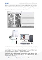
However, technical challenges still remained for precise embolism control in the mechanistic studies of brain damage and repair after perinatal arterial ischemic stroke (PAIS). LSCI provided a measure of blood flow velocity by quantifying the extent of blurring of dynamic speckles caused by the motion of red blood cells through the vessels. As vindicated by our client, “the traditional pathological sectioning methods and neurological assessment tools are unable to accommodate the needs of the MCAO models. This is because there are disparities between different types of infarcts even though their...
Open the catalog to page 3
[2] B. David,‘Laser Doppler, speckle and related techniques for blood perfusion mapping, Physiological Measurement (2001). [3] L. Yuan, and L. Hongyang, and L. Hangdao, T. Shanbao,‘Photothrombotic Ischemia: A Minimally Invasive and Reproducible Photochemical Cortical Lesion Model for Mouse Stroke Studies’, J Vis Exp (2013) [4] Precise control of embolic stroke with magnetized red blood cells in mice,Communications Biology volume 5, Article number: 136 (2022), https://www.nature.com/articles/s42003-022-03082-9
Open the catalog to page 4Archived catalogs
 RWD Surgical Instruments
18 Pages
 RWD Infusion imager RFLSI Ⅲ
2 Pages
 RWD Rotating impactor 68099Ⅱ
2 Pages
 RWD Syringe Pump R462
1 Page
 Rotary Microtomes S710
2 Pages
 RWD Veterinary medical equipment
32 Pages
 RWD Microcentrifuge M1324R
2 Pages
 RWD Osmotic infusion Pump
2 Pages
 RWD Gradient Thermal Cycler
2 Pages
 2015 Product Catalogue
44 Pages
 PRODUCT CATALOGUE 2017
51 Pages


- Medical dolly
- Detection kit
- Logistics trolley
- SAI Infusion surgery forceps
- SAI Infusion grasping forceps
- Microscopy
- Medical kit
- Laboratory incubator
- Compound microscope
- Valve
- Cannula
- Laboratory centrifuge
- Patient monitor
- Stainless steel forceps
- Laboratory microscope
- Benchtop centrifuge
- Desktop microscope
- Dental treatment unit
- SAI Infusion straight forceps
- SAI Infusion surgery scissors