
- Catalogs
- RWD Life Science
- RWD How to create a successful MCAO Stroke model?


- Company
- Products
- Catalogs
- News & Trends
- Exhibitions
RWD How to create a successful MCAO Stroke model?

RWD How to create a successful MCAO Stroke model?
Stroke is a major cerebrovascular disease with high morbidity, disability, and mortality rates. It is the leading cause of disability and the second leading cause of death. Strokes are categorized into ischemic and hemorrhagic, with ischemic strokes accounting for approximately 80% of cases due to cerebral embolisms.
Creating a Successful MCAO Model
The Middle Cerebral Artery Occlusion (MCAO) model is crucial for studying ischemic strokes. The filament model is one of the methods used to create this model in mice.
Procedure
- Anesthesia: Mice are anesthetized using a specific regime, typically involving Isoflurane, in consultation with veterinary staff.
- Blood Perfusion Check: Laser speckle imaging is used to monitor blood perfusion.
- Surgical Steps: The body temperature of mice is maintained at 36.5°C ± 0.5°C using a heating plate. The skin is disinfected, and a midline neck incision is made. The left common carotid artery (LCCA) is dissected, and ligatures are applied to the LCCA, left external carotid artery (LECA), and left internal carotid artery (LICA).
- Filament Insertion: A monofilament is introduced into the LICA to occlude the origin of the left middle cerebral artery (LMCA). Proper placement is confirmed if blood perfusion declines by more than 70%.
- Post-Operative Care: Mice receive saline for volume replenishment and lidocaine gel for pain relief. If reperfusion is intended, the filament is withdrawn after a set occlusion period, and blood perfusion is rechecked.
Post-Surgery Monitoring
Animals are monitored for body temperature and signs of discomfort. Daily checks are necessary to ensure recovery.
Conclusion
The MCAO filament model is a detailed procedure requiring precise surgical skills and post-operative care to study ischemic strokes effectively.
Catalog excerpts
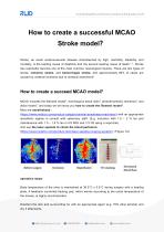
How to create a successful MCAO Stroke model? Stroke, an acute cerebrovascular disease characterized by high, morbidity, disability, and mortality, is the leading cause of disability and the second leading cause of death 1, 5 . Stroke has reportedly become one of the most common neurological injuries. There are two types of stroke, ischemic stroke, and hemorrhagic stroke, and approximately 80% of cases are caused by cerebral ischemia due to cerebral embolisms 6 How to create a succeed MCAO model? MCAO includes the filament model 2 , homologous blood clots 3 , photothrombotic Ischemia 4, and distal model 7, and today we will show you how to create the filament model 2 . Mice are anesthetized : (https://www.rwdstco.com/product-category/animal-anesthesia-machines/) with an appropriate anesthetic regime in consult with veterinary staff. (E.g. induction with 1.5 – 2 % Iso and maintenance with 1.0 – 1.5 % Iso in 2/3 N2O and 1/3 O2 using a vaporizer). And use the laser speckle to check the blood perfusion: (https://www.rwdstco.com/product-item/laser-speckle-imaging-system/). (Figure 1A) operation steps: Body temperature of the mice is maintained at 36.5°C ± 0.5°C during surgery with a heating plate. A feedback controlled heating pad, which warms according to the rectal temperature of the mouse, is highly recommended. Disinfect the skin and surrounding fur with an appropriate agent (e.g. 70% ethyl alcohol) and dry it afterwar
Open the catalog to page 1
A midline neck incision is made and the soft tissues are pulled apart. The left common carotid artery (LCCA) is carefully dissected free from the surrounding nerves (without harming the vagal nerve) and a ligature is made using 6.0/7.0 string. 5.0 string can also be used. The left external carotid artery (LECA) is then separated and a second knot is made. Next, the left internal carotid artery (LICA) is isolated and a knot is prepared with a 6.0 filament. After obtaining good view of the left internal carotid artery (LICA) and the left pterygopalatine artery (LPA), both arteries are clipped,...
Open the catalog to page 2
Figure 2. Scheme of the vessel architecture supplying the brain (depicted in background) in the mouse. Different strains may show variations, for example the occipital artery sometimes leaves from the internal carotid artery. Reference: 1 Y. Dou, Z. Wang, and G. Chen, ‘The Role of Hydrogen Sulfide in Stroke’, Med Gas Res, 6 (2016), 79-84. 2 O. Engel, S. Kolodziej, U. Dirnagl, and V. Prinz, ‘Modeling Stroke in Mice – Middle Cerebral Artery Occlusion with the Filament Model’, J Vis Exp (2011). 3 R. Jin, X. Zhu, and G. Li, ‘Embolic Middle Cerebral Artery Occlusion (Mcao) for Ischemic Stroke with...
Open the catalog to page 3Archived catalogs
 RWD Surgical Instruments
18 Pages
 RWD Infusion imager RFLSI Ⅲ
2 Pages
 RWD Rotating impactor 68099Ⅱ
2 Pages
 RWD Syringe Pump R462
1 Page
 Rotary Microtomes S710
2 Pages
 RWD Veterinary medical equipment
32 Pages
 RWD Microcentrifuge M1324R
2 Pages
 RWD Osmotic infusion Pump
2 Pages
 RWD Gradient Thermal Cycler
2 Pages
 2015 Product Catalogue
44 Pages
 PRODUCT CATALOGUE 2017
51 Pages



- Medical dolly
- Detection kit
- Logistics trolley
- RWD surgery forceps
- RWD grasping forceps
- Laboratory incubator
- Medical kit
- Microscopy
- Cannula
- Laboratory centrifuge
- Compound microscope
- Stainless steel forceps
- Benchtop centrifuge
- Patient monitor
- Laboratory microscope
- Dental treatment unit
- RWD straight forceps
- Cytology reagent
- Desktop microscope
- Blood centrifuge