
- Catalogs
- RWD Life Science
- RWD Infusion imager RFLSI Ⅲ


- Company
- Products
- Catalogs
- News & Trends
- Exhibitions
RWD Infusion imager RFLSI Ⅲ

RWD Infusion imager RFLSI Ⅲ
- Model: RFLSI Ⅲ
- Monitoring Band: 0-5000 PU
- Image Acquisition Speed: Up to 120 FPS
- Image Size: Less than 90mm x 90mm
- Camera: Dual CMOS (Color), 5.0 million pixels (2048 x 2048)
- Zoom Range: 12× optical zoom with auto-focus
- Monitoring Laser: 785 nm, 110mW
- Indicator Laser: 660 nm
- Region of Interest (ROI): Square, circle, polygon
- Quantized Data: Provides in-vivo data and quality control standards for ischemia models.
- Real-time Data: Allows for real-time monitoring of reperfusion data during ischemia models.
- Visual Data: Offers 4K imaging capabilities for detailed visualization of blood vessels.
- Studies on diabetic peripheral neuropathy in mice using DW14006 as a direct AMPK activator.
- Research on platelet activation involving the 14-3-3ζ-c-Src-integrin-β3 complex.
- Investigation of meningeal lymphatic vessels in clearing erythrocytes from subarachnoid hemorrhage.
- Analysis of arterial supercharging benefits on flap survival, showing significant differences in vascular dilation and perfusion values.
Catalog excerpts

Applications Cerebral Blood Flow Monitoring Cortical Spreading Depolarization (CSD) Neovascularization/ Angiogenesis Stroke Model (MCAO) Skin Blood Flow Monitoring Organ blood flow monitoring Chicken Chorioallantoic Membrane (CAM) Assay Hind Limb Ischemia (HLI) Laser Speckle Imaging System F o r P r e - C l i n i c a l R e s e a r c h a b o u t V a s c u l a r. Parameter Model Image acquisition speed Image size Camera Zoom range Monitor camera Image camera Monitoring Laser Laser power Indicator laser Region of interest(ROI) ROI shape Vascular diameter Data Dual CMOS(Color) On-line & Off-line analysis Square, circle, polygon On-line & Off-line analysis Imagine, Video Blood Perfusion RWD Life Science Co.,Ltd Add: 6540 Lusk Blvd, Suite C161 San Diego, CA 92121 U.S.A Te l : (8 5 8 ) 9 0 0 - 5 8 7 9 E-ma il : s e r v i ce@r wdl s .com RWD RFLSI Ⅲ Laser Speckle Imaging System- Flyer-V1.0-2021-02-25-EN
Open the catalog to page 1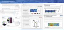
Laser Speckle imaging system (RFLSI Ⅲ) Laser Speckle Imaging System (RFLSI Ⅲ) is a blood perfusion imager based on Laser Speckle Contrast Imaging (LSCI) technology. LSCI provides a better means to study the microcirculation that were not possible in the past. It allows visualization of tissue blood perfusion and imaging with high time and spatial resolution. There is no influence on the perfusion, as no contact to the tissue is needed, nor dyes or tracer elements. Speckle flow techniques are based on the changes over time of the dynamic speckle pattern generated by motion in the sample. In these...
Open the catalog to page 2Archived catalogs
 RWD Surgical Instruments
18 Pages
 RWD Rotating impactor 68099Ⅱ
2 Pages
 RWD Syringe Pump R462
1 Page
 Rotary Microtomes S710
2 Pages
 RWD Veterinary medical equipment
32 Pages
 RWD Microcentrifuge M1324R
2 Pages
 RWD Osmotic infusion Pump
2 Pages
 RWD Gradient Thermal Cycler
2 Pages
 2015 Product Catalogue
44 Pages
 PRODUCT CATALOGUE 2017
51 Pages



- Medical dolly
- Detection kit
- Logistics trolley
- RWD surgery forceps
- RWD grasping forceps
- Laboratory incubator
- Medical kit
- Microscopy
- Cannula
- Laboratory centrifuge
- Compound microscope
- Stainless steel forceps
- Benchtop centrifuge
- Patient monitor
- Laboratory microscope
- Dental treatment unit
- RWD straight forceps
- Cytology reagent
- Desktop microscope
- Blood centrifuge