- Catalogs
- RWD Life Science
- RWD Methods to improve the success rate of MCAO model construction
- Company
- Products
- Catalogs
- News & Trends
- Exhibitions
RWD Methods to improve the success rate of MCAO model construction
1 /4Pages

RWD Methods to improve the success rate of MCAO model construction
1 /4Pages
Catalog excerpts
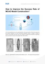
How to Improve the Success Rate of MCAO Model Construction? (1)Fix the anesthetized rat on the operating table in the supine position. Shave the surgical site and conduct disinfection. Use the conventional ophthalmic scissor or scalpel to create an incision at the middle of the neck, then use a tweezer to perform blunt dissection of glandular tissue and fascia to expose and separate CCA, ECA and ICA. Aslan et al., Experimental & Translational Stroke Medicine, 2014 (“The image has been grayscaled”
Open the catalog to page 1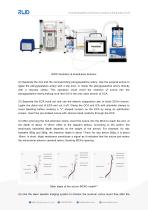
RWD Ventilator & Anesthesia Solution (2) Separate the ICA and the corresponding pterygopalatine artery. Use the surgical suture to ligate the pterygopalatine artery with a slip knot, or clamp the pterygopalatine artery directly with a vascular clamp. This operation could avoid the insertion of suture into the pterygopalatine artery,making sure that ICA is the only open branch of CCA. (3) Separate the ECA trunk out and use the electric coagulation pen to block ECA’s branch. Ligate the distal end of ECA and cut it off. Clamp the CCA and ICA with arteriolar clamps to avoid bleeding before creating...
Open the catalog to page 2
surgery. 70%~80% decreases of the blood flow indicate the successful establishment of the model. (6) Stitch the subcutaneous tissue and skin after ligation and disinfection. Put the rat back into the cage after it recovers from anesthesia. (7) Only by pulling out the suture to allow the silicone head to return to the ECA, the MCA’s blood flow can be restored, as blood flow from the CCA can be reperfusion into the MCA. (8) The control group model almost follows the same procedure, while the only difference is that the suture is not inserted after vessel separation, and subcutaneous tissue and...
Open the catalog to page 3
exclusion criteria. Focal ischemic stroke in animals is typically induced by occlusion of the middle cerebral artery. However, the models of middle cerebral artery occlusion including the suture and embolic methods are imperfect in causing a sustained reduction in blood flow. It is possible in some situations that occlusion may occur but spontaneous reperfusion may ensue, leading to infarct size variability. Basic physiological parameters such as blood pressure, temperature, blood gases, and blood glucose should be routinely monitored. Temperature should be maintained within the normal physiological...
Open the catalog to page 4Archived catalogs
 RWD Surgical Instruments
18 Pages
 RWD Infusion imager RFLSI Ⅲ
2 Pages
 RWD Rotating impactor 68099Ⅱ
2 Pages
 RWD Syringe Pump R462
1 Page
 Rotary Microtomes S710
2 Pages
 RWD Veterinary medical equipment
32 Pages
 RWD Microcentrifuge M1324R
2 Pages
 RWD Osmotic infusion Pump
2 Pages
 RWD Gradient Thermal Cycler
2 Pages
 2015 Product Catalogue
44 Pages
 PRODUCT CATALOGUE 2017
51 Pages


- Medical dolly
- Detection kit
- Logistics trolley
- SAI Infusion surgery forceps
- SAI Infusion grasping forceps
- Microscopy
- Medical kit
- Laboratory incubator
- Compound microscope
- Valve
- Cannula
- Laboratory centrifuge
- Patient monitor
- Stainless steel forceps
- Laboratory microscope
- Benchtop centrifuge
- Desktop microscope
- Dental treatment unit
- SAI Infusion straight forceps
- SAI Infusion surgery scissors