- Catalogs
- RWD Life Science
- RWD Used the Laser Speckle Imaging System to monitor the blood perfusion for Hind Limb Ischemic (HLI)
- Company
- Products
- Catalogs
- News & Trends
- Exhibitions
RWD Used the Laser Speckle Imaging System to monitor the blood perfusion for Hind Limb Ischemic (HLI)
1 /2Pages

RWD Used the Laser Speckle Imaging System to monitor the blood perfusion for Hind Limb Ischemic (HLI)
1 /2Pages
Catalog excerpts
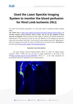
Used the Laser Speckle Imaging System to monitor the blood perfusion for Hind Limb Ischemic (HLI) The rodent limb ischemia preparation is a wild used model of peripheral arterial disease (PAD). The model uses a https://www.rwdstco.com/product-item/laser-speckle-imaging-system/ to real-time measure blood perfusion (PU) in limbs, and we can get imaging of blood vascular to distribute. When we draw the ROIs (region of interest), the software enables the measurement of the blood flow. As we can see, the difference in PU between the ischemic versus and non-ischemic limb (Fig1). And we can do some research about the “Reperfusions” which can measure the function of drugs in the PAD. And if you want to have a free demo of the laser speckle,http://online.rwdstco.com/laserspeckleimage . Equipment recommendations The Laser Speckle Contrast Imaging System (RFLSI Ⅲ ) offers the highest spatial resolution (2048 × 2048) and can be used to assess reperfusion in the pads of the feet(https://pubmed.ncbi.nlm.nih.gov/32647036/). RFLSI recording the process of reperfusions and vessel formation
Open the catalog to page 1
Fig 1. Right limb: ischemic versus, left limb: non-ischemic limb. Reference: https://pubmed.ncbi.nlm.nih.gov/32647036/
Open the catalog to page 2Archived catalogs
 RWD Surgical Instruments
18 Pages
 RWD Infusion imager RFLSI Ⅲ
2 Pages
 RWD Rotating impactor 68099Ⅱ
2 Pages
 RWD Syringe Pump R462
1 Page
 Rotary Microtomes S710
2 Pages
 RWD Veterinary medical equipment
32 Pages
 RWD Microcentrifuge M1324R
2 Pages
 RWD Osmotic infusion Pump
2 Pages
 RWD Gradient Thermal Cycler
2 Pages
 2015 Product Catalogue
44 Pages
 PRODUCT CATALOGUE 2017
51 Pages


- Medical dolly
- Detection kit
- Logistics trolley
- SAI Infusion surgery forceps
- SAI Infusion grasping forceps
- Microscopy
- Medical kit
- Laboratory incubator
- Compound microscope
- Valve
- Cannula
- Laboratory centrifuge
- Patient monitor
- Stainless steel forceps
- Laboratory microscope
- Benchtop centrifuge
- Desktop microscope
- Dental treatment unit
- SAI Infusion straight forceps
- SAI Infusion surgery scissors