- Catalogs
- SBM Sistemi
- OS1000
OS1000
1 /16Pages

OS1000
1 /16Pages
Catalog excerpts
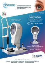
Embrace the power ofTWO over ONE! With OSIOOO approaching Dry Eye analysis has never been easier. More exams, one device. Lift the unique AUTO INTERFEROMETRY PANEL setting a new standard in precision diagnostics. Revolutionize Dry Eye Care: Unleash comprehensive assessment with our Dedicated Platform, aligned with TFOS DEWS II Recommendations
Open the catalog to page 1
Unique technology for automatic and objective analysis of patients with MGD Tear film interferometry is increasingly being used in research to observe the tear film. Interferometry is a technique that studies the surface refractive pattern and dynamics of the lipid layer of the tear film, thus allowing measurement of tear film stability and lipid layer thickness. Interferometers are investigative tools used in many fields of science and engineering. They are called interferometers because they work by combining two or more light sources to create an interference pattern, which can be measured...
Open the catalog to page 2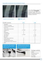
An A.I. based algorithm upscales the acquired image to 23 Megapixels. The extreme quality image is then elaborated obtaining new levels of precision in Placido disk based corneal topography measurement. OSIOOO versions plus full Joystick One-click acquisition Images and movies can be captured instantly and conveniently by pressing the joystick button. Left/Right automatic detection OSIOOO automatically recognizes the right and left eye, allowing an even faster diagnosis of the ocular surface. Package contents OSIOOO Base Plate and Chinrest Calibration sphere ICP Software Power supply
Open the catalog to page 3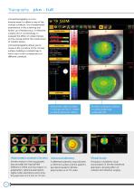
Corneal topography is a non-invasive exam to obtain a map of the corneal curvature. It is a fundamental examination in the screening and follow-up of keratoconus, in refractive surgery and in contactology, to evaluate the effect of contact lenses on the cornea and for the construction of contact lenses. Corneal topography allows you to measure the curvature of the corneal surface, building a colored map in which each color corresponds to a different curvature. Acquisition Quality Summary indices Horizonta Visib e ris Diameter Pupil (Topographic) . haoe ndices Keratometric data including K-readings...
Open the catalog to page 4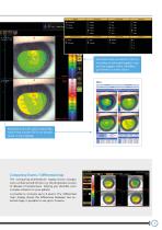
Klyce/Wil*on Schwind Comparing Exams / Differential map The "comparing examinations" display shows changes over a certain period of time, e.g. the progressive course of disease of keratoconus. helping you describe even complex situation to your patient. Is possible to compare up to 4 exams. The "differential map" display shows the differences between two selected maps, is possible to use up to 3 exams.
Open the catalog to page 5
The auto fit module combine the topographic data and RGP lens data, to find and fit the best solution for the patient's eye, simulating the fitting with fluoresceine. With OSIOOO is possible to acquire in vivo fluoresceine image of the lens or testing the fitting with simulated fluorescein visualization. The contact lens simulation produces an image of how a specific lens fits the eye. The simulation allows you to adjust the angle and position of the contact lens and includes automatic recalculation of the fluorescent image. The system allows you to order fewer lenses and reduce chair time while...
Open the catalog to page 6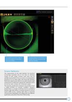
Simulated fluorescein image to verify the distance of the lens from the cornea. Cutaway graph of the distance of the lens from the cornea on the selected meridian. The measurement of the pupil diameter has become increasingly important also in the field of refractive surgery as well. Larger scotopic pupil sizes may be partially responsible for the occurrence of postoperative symptoms such as halos, glare, and monocular diplopia. Refractive surgeons also need an accurate scotopic pupil measurement to determine appropriate treatment zones for excimer laser, corneal, and intraocular surgery. The...
Open the catalog to page 7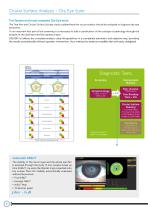
The fastest and most complete Dry Eye suite The Tear film and Ocular Surface Society study outlined how the ocular surface should be analyzed to diagnose dry eye syndrome. To an important first part of the screening it is necessary to add a classification of the subtype of pathology through the analysis of the lipid layer and the aqueous layer. OSIOOO Full allows the complete analysis using the guidelines in a completely automatic and objective way, providing the results automatically without operator intervention, thus making the analysis incredibly fast and easily delegated. Diagnostic Tests...
Open the catalog to page 8
Interferometry Thanks to the anterior illumination module, OSIOOO can aquire the lipid layer secrection on the cornea. The device highlights the lipid layer and the software evaluates the quantity and quality of the lipid component on the tear film, plus subjective • full automatic Subtype Classification Tests Aqueous deficiency • Low volume MGD means Meibomian Glands Disfunction This condition happens when the meibomian glands are not working as needed. To verify this condition a simple Meibography is not enough to know the working condition of the patient's glands. T Automatic tear meniscus...
Open the catalog to page 9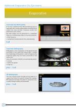
Additional Evaporative Dry Eye exams Automatic Eye blink quality It has been established that efficient blinking plays an important role in ocular surface health including during contact lens wear and that it improves contact lens performance and comfort. Eye blink analysis can be performed on a dedicated video or on interferometry video to know automatically: • Blink frequency • Partial blink (Fundamental for MG understanding) full Automatic Meibography Meibography is the visualization of the glands through illumination of the eyelid with infrared light. It images the morphology of the glands...
Open the catalog to page 10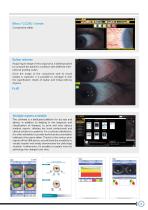
Bulbar redness Acquiring an image of the conjunctiva, it will be possible to compare the patient's condition with different international grading scales. Once the image of the conjunctiva with its blood vessels is captured, it is possible to compare it with the classification sheets of bulbar and limbal redness degrees, full Multiple reports available The software is a dedicated platform for dry eye and allows, in addition to helping in the diagnosis and classification of diseases, to print and save various medical reports, offering the most professional and clinical solutions to patients. For...
Open the catalog to page 11


- Medical support arm
- Tablet computer software
- Tablet PC software
- Windows software
- Fixed ophthalmic examination
- Measurement software
- IPL system
- Desk support arm
- Slit lamp
- Table slit lamp
- Table-top IPL system
- Tablet PC support arm
- Ophthalmic software
- Digital slit lamp
- Android application
- Eye software
- Eye chart
- Corneal topographer
- IOS iOS application
- Dry eye diagnosis system