
- Catalogs
- Shenzhen Bestman Instrument
- vascular doppler BV-650

vascular doppler BV-650
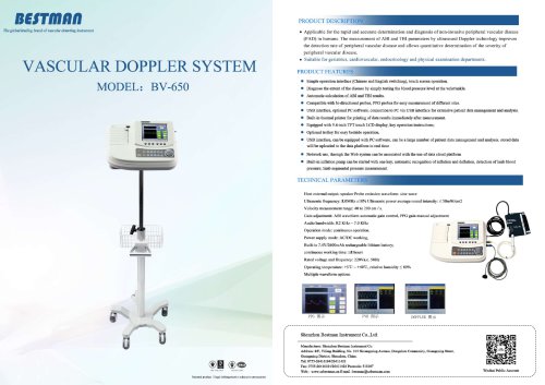
vascular doppler BV-650
The document appears to reference three types of technical illustrations or diagrams: PPG (Photoplethysmogram), PVR (Pulse Volume Recording), and Doppler diagrams. These are commonly used in medical diagnostics to assess vascular and circulatory conditions.
PPG (Photoplethysmogram) Diagram
PPG diagrams illustrate blood volume changes in the microvascular bed of tissue, typically used to monitor heart rate and blood flow. Key points include waveform shape, amplitude, and timing, which help in evaluating peripheral circulation.
PVR (Pulse Volume Recording) Diagram
PVR diagrams represent pulse volume changes in limbs, useful for detecting arterial occlusions or peripheral artery disease. Important parameters include pulse amplitude and waveform contour, which indicate blood flow quality and vessel patency.
Doppler Diagram
Doppler diagrams display blood flow velocity and direction using ultrasound technology. They are critical for assessing vascular stenosis, occlusions, and blood flow dynamics. Key features include spectral waveforms, peak systolic velocity, and flow patterns.
Overall, these diagrams serve as essential tools in vascular diagnostics, each providing complementary information about blood flow and vessel health.
All Shenzhen Bestman Instrument catalogs and technical brochures
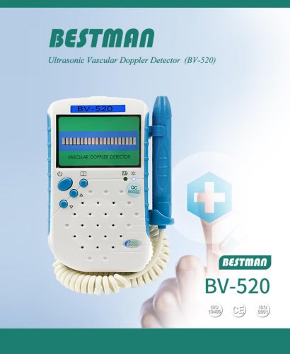 BV-520 portable vascular dopplers
13 Pages
 Bestman company catalogue 2025
38 Pages
 ShenZhen Bestman Instrument
66 Pages
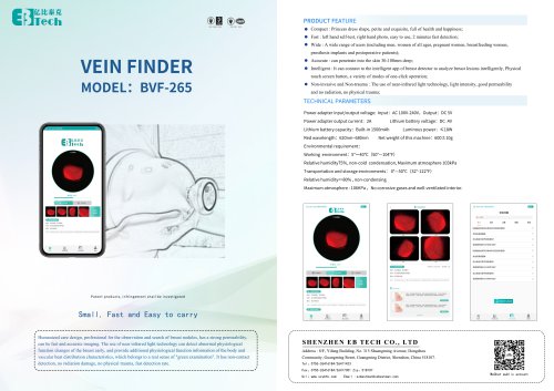
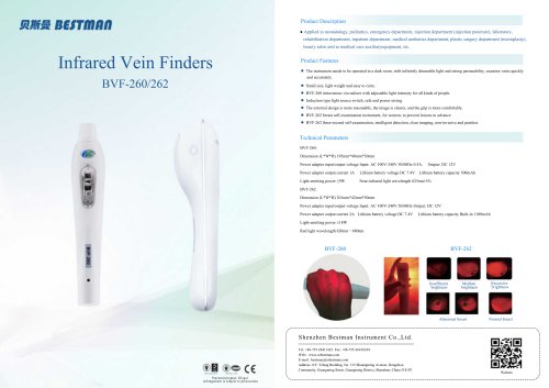 vein finders BVF-260
1 Page
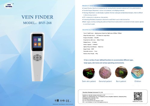
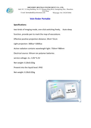 vein finder BVF-263
6 Pages
 syringe destroyer catalogue
13 Pages
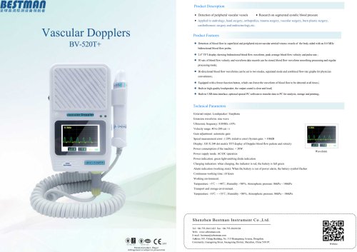
 Vascular Doppler Catalogue
19 Pages
 2024 new catalogue
20 Pages

Archived catalogs
 BSM Catalogue 2021
20 Pages
- Laboratory incubator
- Thermometer
- Medical thermometer
- SpO2 monitor
- Temperature-control laboratory incubator
- Medical pulse oximeter
- EKG
- Digital electrocardiograph
- Fingertip pulse oximeter
- Infrared thermometer
- Forehead thermometer
- Electrocardiograph with screen
- Electrocardiograph with printer
- Non-contact thermometer
- Floor-standing laboratory incubator
- Hand-held pulse oximeter
- Fetal doppler
- Pocket doppler
- Wired electrocardiograph