Catalog excerpts
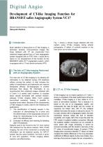
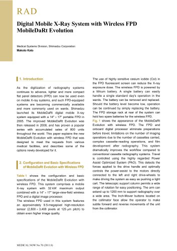
Digital Angio Development of CT-like Imaging Function for BRANSIST safire Angiography System VC17 Medical Systems Division, Shimadzu Corporation Kazuyoshi Nishino 1. Introduction Much attention is being given to CT-like imaging, a technique whereby cross-sectional images like those obtained with CT are constructed from rotational images gained from a C-arm angiography system equipped with an FPD. Here, I would like to report on our development of this function for the BRANSIST safire VC17 angiography system, which is equipped with a 17-inch direct-conversion FPD. Fig. 1 shows a clinical image obtained with this system using CT-like imaging during arterial portography. A defect of contrast medium in the liver tumor can be seen clearly. 2. The Aim of CT-like Imaging Performed with an Angiography System The main aim of CT-like imaging is, by allowing 3D information to be obtained during IVR treatment without moving the patient, to help increase the safety of examinations and treatments and improve therapeutic efficacy. 3D-angiography is another technique that allows 3D information to be reconstructed from rotational images obtained with a C-arm. It is mainly used, however, to visualize blood vessels illuminated with contrast medium, whereas CT-like imaging offers much greater low-contrast resolution, making it possible to visualize tumor stains. Because of this characteristic, CT-like imaging is mainly used for the following two applications: 1) The identification of feeding vessels in the execution of chemotherapy for a tumor and the determination of the appropriate amount of drugs for chemotherapy to be used from the size of the affected area. 2) Evaluation of efficacy and checking for bleeding after IVR treatment. Patients used to be moved to the CT room during IVR treatment for the above reasons. With CT-like imaging, the risk of this movement is avoided and information can be obtained quickly. An IVR-CT system has the same functions, but both in terms of cost and operation, CT-like imaging attains superior cost-effectiveness. 3. CT vs. CT-like Imaging CT-like imaging is by no means superior to CT. Table 1 shows a comparison of the basic performance of the two modalities. CT-like imaging is, in fact, inferior to CT in terms of low-contrast resolution. This is because it is based on the use of an angiography system, and consequently the data bit length and the number of exposures are limited. Also, with CT, the scattered radiation that enters the sensor is reduced to around 1% by a grid, whereas with an angiography system, the basic mechanical structure makes this kind of reduction impossible, and roughly half of the X-rays that enter the FPD consist of scattered radiation component. Although this scattered radiation component can be corrected to some extent with image processing, it cannot be removed accurately, and this results in inaccuracy of the CT values of reconstructed images. At present, CT-like imaging does not produce absolute CT values, but rather visualizes the relative differences in the X-ray absorption coefficients of different objects. As the name implies, it is a technique that produces images that are comparable to, MEDICAL NOW No.64 (2008.7)
Open the catalog to page 1
but not the same as, those produced with CT. On the other hand, CT-like imaging is superior to CT in some aspects. One such aspect is high-contrast resolution (spatial resolution). With CT-like imaging, it is possible to create isotropic 3D images with a high level of spatial resolution. Also, the system that we developed can perform acquisition for CT-like imaging with an exposure level of no greater than one fifth to one half that of CT. This yields clinical benefits. For example, in cases where there are multiple feeding vessels, CT-like imaging is acceptable for each one. CT-like...
Open the catalog to page 2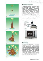
The BRANSIST safire VC17 angiography system is shown in Fig. 4. We developed a CT-like imaging function (including a 3D-angiography function) as an option for this system (Fig. 5). The image processing workstation is connected to the imaging system via high-speed LAN, and after rotational images are acquired, reconstructed images can be displayed in as little as 90 sec. A CT viewer is used for image observation, and in addition to the 2D display of axial images, MPR, CPR (curved MPR), MIP, volume rendering (VR), and virtual endoscopy (VE) are supported. Various types of masking, the...
Open the catalog to page 3Archived catalogs
-
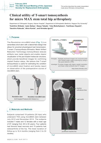
 Tomosynthesis
6 Pages
-
 FLEXAVISION F3
3 Pages
-
 RADSpeed Pro
4 Pages
-
 DynamicStentView
4 Pages
-
 X-Ray System
3 Pages
-
 Digital Angio
4 Pages
-
 c12 MIX
4 Pages
-
 RAD
RAD4 Pages
-
 t-smart
2 Pages
-
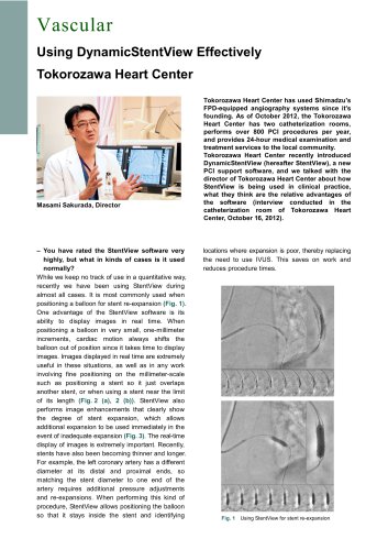 Vascular_interview
3 Pages
-
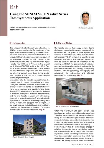 SONIALVSION
4 Pages
-
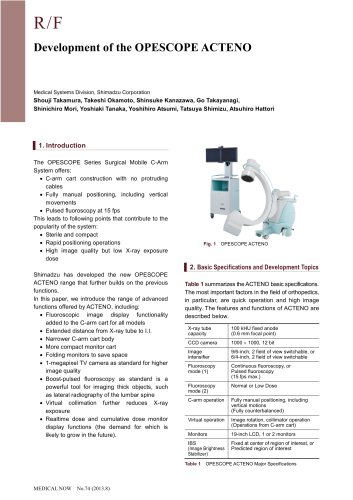 OPESCOPE ACTENO
3 Pages