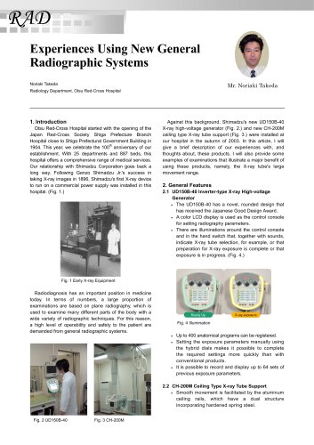
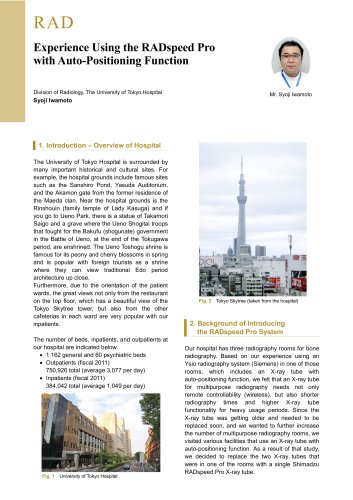
Catalog excerpts

Vascular DynamicStentView: Cardiovascular Intervention Support Software Medical Systems Division, Shimadzu Corporation Yoshiaki Miura Percutaneous coronary intervention (PCI) using angiography systems has become more widespread in recent years, while the associated technologies have become increasingly sophisticated. There has been a corresponding demand for angiography system functions to support advanced medical treatments. In response to this demand, Shimadzu developed the DynamicStentView cardiovascular intervention support software according to an entirely new concept. The aims, operating principles, and applications of the newly developed DynamicStentView software are described below. 2. Background In many cases of patients with advanced arteriosclerosis, the spread of the angiostenosis often requires placement of a new stent alongside a previously placed stent. In such cases, it is important for the new stent to slightly overlap the existing stent. Since stents have an extremely thin construction and poor visibility in X-ray images, clear confirmation of their position is often difficult. The position of a stent being placed is confirmed using markers placed at either end of the balloon on which the stent is mounted before expansion. The balloon is retrieved after placement of the stent. Since there are no markers present on a previously placed stent, it can be difficult to confirm its position in X-ray images. DynamicStentView was developed to simplify the positioning of new stents by improving the visibility of previously placed stents (Fig. 1). To improve the visibility of a previously placed stent, it is necessary to enhance the weak image signals of the stent through addition/averaging processing. However, since the blood vessel in which the stent is placed moves significantly due to cardiac motion, simply adding and averaging the stent image signals is unable to enhance the image of the stent but instead results in diffusion and blurring of the stent image. Therefore, enhancing the stent in the image requires extraction of the stent and markers in each frame of the video image and adding and averaging on each frame while correcting for image deformation due to cardiac motion. This permits the display of an enhanced image of the stent. By performing this processing on a single frame at up to 30 frames per second (within 33 ms), DynamicStentView displays an enhanced image of the stent in realtime during stent placement, with no delay due to post-processing. DynamicStentView also offers a clear display of the relative positions of the previously placed stent and the markers of the stent to be newly placed. Fig. 2 shows an actual example during application, and Fig. 3 shows the operating principle of DynamicStentView in an actual case. Stent enhanced image Fig. 2 Example Application of DynamicStentView Fig. 1 Stent Placement (Images provided by Heisei Shisenkai Foundation Kokura Memorial Hospital)
Open the catalog to page 1
(3.0 mm diameter, 96.5 µm thick) and a chest phantom manufactured by Kyoto Kagaku that represents a chest thickness of 20 cm. The results of using DynamicStentView were then verified. The experimental set up is shown in Fig. 5. The experiment confirmed enhancement and improved visibility of the stent image. Example after processing three immediately previous frames Fig. 3 Operating Principle of DynamicStentView Ex p a n d e d ste n t 4. The Principle Behind Extracting Markers from an Image The most important function for DynamicStentView is extracting the markers to use as the reference for...
Open the catalog to page 2
6. Operability (screen description) This section describes DynamicStentView by focusing on its operation. DynamicStentView is useful for confirming positioning when using a balloon to re-expand a stent, or when placing overlapping stents. Fig. 6 shows examples of the clinical applications of DynamicStentView. Using a balloon to re-expand a stent B a llo o n fo r r e -e x p a n sio n Placing overlapping stents B a llo o n a tta c h e d to th e ste n t to b e in se r te d Fig. 6 Clinical Applications of DynamicStentView Operation is simple. Press a button on the bedside console to select...
Open the catalog to page 3
(3) DetectingView area Appropriate marker detection is important for enhancement processing. The marker extracted from each frame of the video image is displayed in the DetectingView area to confirm whether markers are detected correctly. If something other than the marker is recognized by mistake, the mistakenly recognized object is then displayed. While viewing the DetectingView area display, the collimator can be used to remove the mistakenly recognized object from the field of view to ensure appropriate marker detection. confirmation. Subsequently, it is not necessary to cancel the...
Open the catalog to page 4Archived catalogs
-
 Tomosynthesis
6 Pages
-
 FLEXAVISION F3
3 Pages
-
 RADSpeed Pro
4 Pages
-
 X-Ray System
3 Pages
-
 Digital Angio
4 Pages
-
 c12 MIX
4 Pages
-
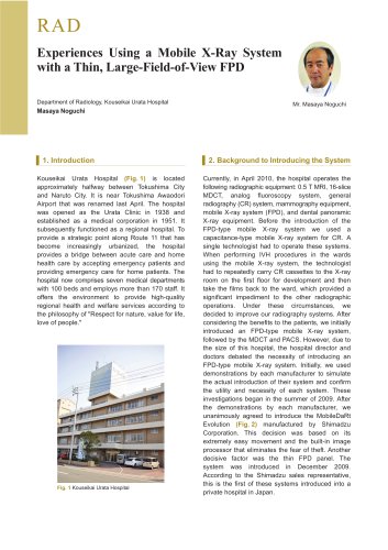 RAD
RAD4 Pages
-
 t-smart
2 Pages
-
 Vascular_interview
3 Pages
-
 SONIALVSION
4 Pages
-
 CT-like
3 Pages
-
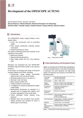 OPESCOPE ACTENO
3 Pages