
Catalog excerpts

White Paper ShearWave™ Elastography Jeremy Bercoff, PhD SuperSonic Imagine Aix en Provence, France Copyright 2008 SuperSonic Imagine S.A. All rights reserved
Open the catalog to page 1
Introduction One of the key medical methods for detection and stiffness (or elasticity). It provides an imaging representation of characterization of pathologies is the assessment of tissue stiffness what was historically assessed qualitatively by palpation. The main by palpation. The importance of clinical assessment of tissue objectives of elastography are to improve diagnostic confidence stiffness has been known since ancient times: In Pharonic Egypt, and increase specificity of the ultrasound exam. more than 5000 years ago, physicians practiced palpation of body parts to determine...
Open the catalog to page 2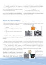
There are two types of mechanically induced waves: far in medical imaging despite the fact that they are explicitly related • to tissue stiffness. In fact, elasticity (E) and shear wave propagation Compression (or bulk) waves: These propagate very quickly in tissue (1500m/s) by successively compressing tissue speed (c) are directly linked through the simple formula: E = 3ρc² (2) layers. Echoes of compressional waves on tissue scatterers are used to perform standard ultrasound imaging. • where ρ is the density of tissue expressed in kg/m3. Shear waves: These are much slower than...
Open the catalog to page 3
• Shear wave based elastography makes use of transient The SuperSonic Imagine Aixplorer™ is the first ultrasound pulses to generate shear waves in the body [14-16]. The tissue’s system to leverage this technology and implement a true shear elasticity is directly deduced by measuring the speed of wave wave based elastography imaging concept. propagation as indicated in formula (2). Shear wave based elastography is the only approach able to provide quantitative and local elastic information in real time [17]. However, its implementation as an imaging mode requires major technological...
Open the catalog to page 4
Shear Wave Generation Shear waves can be generated in the body in different ways. The beating heart is a natural source of shear waves but its vibrations remain localized in its vicinity. The use of external vibrators, such as those used in dynamic MR elastography, are not ideal in the ultrasound environment as they require the manipulation of two devices simutaneously [20]. ShearWave™ Elastography uses the acoustic radiation force induced by ultrasound beams to perturb underlying tissues. This pressure or “acoustic wind” pushes the tissue in the direction of propagation. An elastic medium...
Open the catalog to page 5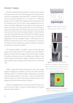
Ultrafast™ Imaging The shear waves generated using the SonicTouch™ excitation need to be captured by the ultrasound system. Shear waves typically propagate in tissues at speeds between 1 and 10 m/s (corresponding to tissue elasticity from 1 to 300 kPa). Consequently, they cross an ultrasound image plane of 3 to 6 cm width in 10 - 20 milliseconds (less than 1/50 of a second). Modern radiology ultrasound systems generate only 50 - 60 images per second. This is too slow to image a propagating shear wave since the shear wave will have disappeared in the time needed to make a single frame. In...
Open the catalog to page 6
Elasticity Estimation To compute a full elasticity image as displayed on the system screen (Figure 5), several supersonic lines are generated using SonicTouch™ Technology, as illustrated on Figure 13. For each line, several ultrafast images are acquired and the shear wave propagation velocity movie is computed. Shear wave speed maps from all the pushing lines are calculated and then combined into a final image. The elasticity map in kPa is directly derived from the final speed map using equation (2). Figure 13 : A small number of supersonic lines generate a full SonicTouch™ technology...
Open the catalog to page 7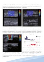
The fibroadenoma in Figure 15 exhibits a mean low elasticity Figure 17 shows an example of two small cysts which appear as value (mean 28 kPa) but has slightly harder contours that can be black voids on the elasticity image, indicating a high probability of seen both on the ultrasound and elasticity image (mean 40 kPa). liquid content. Figure 17 : SWE on two small cysts. Figure 15 : Elasticity map of a fibroadenoma. The mean elastic value of the lesion is 28 kPa. was classified as ACR4 using the BI-RADS® lexicon [23], appears ShearWave™ Elastography in order to evaluate the quantitative as...
Open the catalog to page 8
The quantitative estimation of tissue elasticity offers SWE can help the radiologist to better understand tissue and previously unavailable information that could be incorporated pathologic morphology. The advantages of a local depiction of into the diagnostic decision making process. elasticity are shown in Figure 20. This lesion, classified as ACR5 using ultrasound BI-RADS® lexicon, shows a hypoechoic center and heterogeneous borders on B-mode ultrasound. Local estimation opens new perspective to lesion analysis The local values of the elasticity measurements is one of the main benefits...
Open the catalog to page 9
Real-time operation improves scanning workflow With static elastography systems, tissue strain is displayed in realtime but the elasticity information needs to be extracted by the user using a precise protocol. Typically, a set of real-time strain images is acquired while the radiologist manually vibrates the tissue. The best representation of the tissue’s elastic properties. An indication of the quality of the strain image is usually provided to help the radiologist, but the choice ultimately depends on a subjective Figure 21 : A series of slices through a spherical harder “lesion”...
Open the catalog to page 10
References [1] Sarvazyan, A.P. (2001). Elastic Properties of Soft Tissue. Handbook of Elastic Properties of Solids, Liquids, and Gases. 3: 107127. [15] Nightingale KR, McAleavey SA, Trahey G. E. Shear wave generation using acoustic radiation force: In vivo and ex vivo results. Ultrasound Med Biol 2003;29(2):1715–1723 [2] Skovoroda et al., (1995). Quantitative analysis of the mechanical characteristics of pathologically changed soft biological tissues. Biophysics, 40(6)1359-1364. [16] Bercoff J, Tanter M, Fink M. Supersonic shear imaging: A new technique for soft tissues elasticity mapping....
Open the catalog to page 11