- Catalogs
- taberna pro medicum
- DUB Brochure
DUB Brochure

DUB Brochure
The DUB SkinScanner systems are high-frequency and high-resolution ultrasound diagnostic tools used in dermatology, cosmetics, and pharmaceutical research. These systems offer various models and frequency ranges to meet diverse requirements, with the 22 MHz ultrasound being a common method for non-invasive skin analysis.
History and Development
Since 1986, the DUB systems have evolved from the first A/B digital ultrasound system designed for dermatology to advanced models with USB connectivity and compatibility with Windows operating systems. The product line includes devices with frequencies ranging from 18 MHz to 100 MHz, offering resolutions from 88 µm to 16 µm.
Technical Specifications
The main units vary in dimensions, weight, and power supply, with sampling rates up to 250 MHz and lateral step widths of 33 µm. They connect via USB 2.0/3.0 and are compatible with Windows XP to Windows 8. Key features include automatic skin thickness and density measurements, multiple viewing modes, and personalizable settings.
Key Features and Applicators
The systems support various linear B-Scan applicators with frequencies up to 75 MHz, providing different penetration depths and resolutions. The devices are CE and FDA 510K certified, ensuring medical compliance.
Applications
The DUB SkinScanner is used for monitoring skin aging, elasticity, and treatment efficacy. It aids in diagnosing skin damage, lesions, and tumor depth, and is applicable in cosmetic research and aesthetic treatments.
Advanced Options
Additional features include the Vacu Elasto Pump for skin elasticity measurement, volume measurement with X-Scan, and early osteoporosis risk detection. The system also supports epiluminescence microscopy for detailed skin analysis.
Conclusion
The DUB SkinScanner systems provide comprehensive solutions for high-frequency ultrasound imaging, offering flexibility, precision, and compatibility with modern computing systems, making them ideal for medical and cosmetic applications.
Catalog excerpts

high frequency ultrasound in medicine and cosmetics
Open the catalog to page 1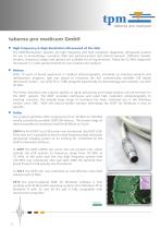
taberna pro medicum GmbH < High Frequency & High Resolution Ultrasound of the Skin The DUB®SkinScanner systems are high frequency and high resolution diagnostic ultrasound systems for use in dermatology, cosmetics field and pharmaceutical and clinical research. Different models, versions, frequency ranges and options are available for all requirements. Today the 22 MHz diagnostic ultrasound is a wide spread method for non-invasive skin analysis. < History After 10 years of broad experience in medical ultrasonography, including an intensive research and development program, tpm was proud to introduce...
Open the catalog to page 2
The entry level in high frequency ultrasound imaging systems. with 22-28 MHz linear B-Scan applicator A cost-effective device ideal for practicing doctors. The DUB®cutis fulfils all requirements for German health insurance. < Key features Ÿ Max. axial resolution: 57 µm at 28 MHz Ÿ Max. digitizing depth: 8 mm Ÿ Scan width: 12.8 mm linear (33 µm step width) Ÿ Medical CE and FDA 510K Ÿ Connection: USB 2.0 or USB 3.0 Ÿ DUB SkinScanner software compatible with all Windows operating systems from XP to Windows 8 Ÿ Viewing modes: B-Scan, A-Scan, Sum-A, ScanLoop (20) Ÿ 2 color scales Ÿ Measurement: length,...
Open the catalog to page 3
The standard system in high frequency ultrasound imaging systems. for 22-28 or 33-38 MHz linear B-Scan applicators The standard device for more flexibility. < Key features Ÿ Max. axial resolution: 42 µm at 38 MHz Ÿ Max. digitizing depth: 16 mm Ÿ Scan width: 12.8 mm linear (33 µm step width) Ÿ Medical CE and FDA 510K Ÿ Connection: USB 2.0 or USB 3.0 Ÿ DUB SkinScanner software compatible with all Windows operating systems from XP to Windows 8 Ÿ Viewing modes: B-Scan, RF-Mode, A-Scan, Sum-A, ScanLoop (2000) Ÿ Filter: Hilbert transformation Ÿ 7 color scales Ÿ Measurement: length, area, density, width,...
Open the catalog to page 4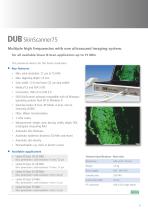
Multiple high frequencies with one ultrasound imaging system. for all available linear B-Scan applicators up to 75 MHz The premium device for the finest resolution. < Key features Ÿ Max. axial resolution: 21 µm at 75 MHz Ÿ Max. digitizing depth: 16 mm Ÿ Scan width: 12.8 mm linear (33 µm step width) Ÿ Medical CE and FDA 510K Ÿ Connection: USB 2.0 or USB 3.0 Ÿ DUB SkinScanner software compatible with all Windows operating systems from XP to Windows 8 Ÿ Viewing modes: B-Scan, RF-Mode, A-Scan, Sum-A, ScanLoop (2000) Ÿ Filter: Hilbert transformation Ÿ 7 color scales Ÿ Measurement: length, area, density,...
Open the catalog to page 5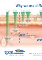
eMail: info@tpm-i
Open the catalog to page 6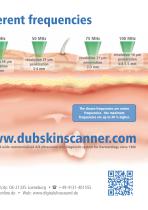
erent frequencies The shown frequencies are center frequencies - the maximum frequencies are up to 40 % higher. d-wide commercialized A/B ultrasound skin diagnostic system for Dermatology since 1986 /city: DE-21335 Lueneburg • ( +49-4131-401555 online.de • Web: www.digitalultrasound.de
Open the catalog to page 7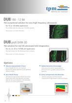
The exceptional solution for very high frequency ultrasound. for 75 or 100 MHz applicators The world-wide one-and-only 100 MHz system! Please contact us for further information. The solution for real 3D ultrasound skin diagnostics. for 22, 33, 50 or 75 MHz 3D-applicators The best choice for 3D ultrasound skin scanners. Please contact us for further information. Options < Volume measurement X-Scan Easy-to-handle with two cross-sectional B-Scans with special applicator tip and additional software module. < Vacu Elasto Pump < Early osteoporosis risk detection Skin elasticity measurement with the...
Open the catalog to page 8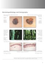
Dermatopathology and Sonography Poorly circumscribed, reddish-brownish tumor on the left leg Poorly circumscribed, asymmetrical, pigmented tumor on the upper back Tentative diagnosis: Malignant melanoma Tentative diagnosis: Malignant melanoma Epiluminescence picture of a tumor Echolucent area with several tissue echos within the whole structure of the poorly differentiated tumor Echolucent area with small echogenic band in the upper part and hypoechoic sharp edged area Dermis and subcutis: Normal echogenicity Dermis and subcutis: Normal echogenicity 22 MHz-Sonogram with DUB SkinScanner Final...
Open the catalog to page 9
Fields of application < Monitoring - skin aging - Mohs surgery - skin elasticity - skin treatment < Diagnosis - tissue beneath wounds and skin Foreign body in scar tissue - 75 MHz - skin damage caused by sun exposure - skin lesions caused by different clinical reasons < Efficacy - laser treatment - wound treatment - cosmetic research - aesthectic skin treatment Before treatment < Detection - skin thickness - osteoporosis risk - tumor depth before and after surgery After 12 months Follow-up after fractional laser photothermolysis
Open the catalog to page 10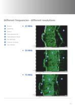
Different frequencies - different resolutions 1 Subcutaneous Tissue Stratum Corneum Hair Follicles
Open the catalog to page 11
Scan viewing modes < A-Scan A complete ultrasound picture is assembled together out of many single A-Scan lines. With the DUB®SkinScanner software it is possible to view all single A-lines. Together with the Sum-A functionality the skin thickness can easily be measured. < B-Scan A complete B-Scan is built up from 384/768 A-Scan lines. Converted into one of 7 available color scales and 2 different transformations (Hilbert and Full-Wave) a typical high frequency ultrasound image for clinical evaluation is created. A-Scan (top) B-Scan (below) view < RF-Scan The RF-Scan shows the original raw data...
Open the catalog to page 12All Taberna pro medicum catalogs and technical brochures
 DUB brochure
12 Pages
 OPFA
OPFA2 Pages
 Dermatology
2 Pages
 Aesthetics
2 Pages